 Credit: Thinkstock
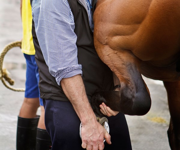
For equine practitioners, lameness is a cornerstone of a veterinary practice—and in some cases, the sole pursuit of their medical experience. While soundness evaluations generate income, there is also a cost associated with them, especially when state-of-the-art equipment is involved.
But, as with any good medical practice, a foundation sets the framework for developing a diagnosis. The starting point is always the clinical exam. Wally Liberman, DVM, of Panorama Equine Medical and Surgical Center in Redding, California, has dedicated his practice primarily to diagnosing and managing lameness concerns. “One of the most important elements to tackling lameness problems is a quality clinical orthopedic exam that involves the power of observation in both static and dynamic evaluation, and hands-on palpation skills,” he said.
Part of a thorough exam involves taking a history from the client, or from the seller in the case of a pre-purchase exam. “The key to taking a good history is to ask the same question three different ways, three different times during the course of the exam,” Liberman explained. “A client’s context is different from a practitioner’s, and it is possible that something they thought wasn’t particularly relevant may be very important.”
To achieve the best results, he continued, “A correct venue with decent footing and a proper surface is critical to achieving a proper orthopedic exam. A firm surface with troughs and inclines brings out torque load and concussion load. Then the horse is moved out on a flat, sand-rubber interfaced arena, or [something] comparable.”
These different surfaces help one discern between soft tissue, bone, joint or hoof issues. Such varying surfaces might not be available at all client barns, so a practitioner with a clinic will have an expense associated with setting up specific areas devoted to lameness workups. Liberman also recommended that, when possible, the horse should be examined with (and exercised by) a technician who understands the mechanics of an orthopedic exam (another expense). With a good handler and a specific protocol, a practitioner is able to achieve the most consistent assessment of a gait analysis.
Hands-on palpation skills are important and are only as good as the practitioner, said Liberman. “It is an art form that relies on true knowledge of anatomy to form a three-dimensional concept of what one is evaluating and feeling. This is coupled with the ability of a practitioner to identify, observe and feel what he or she is evaluating.”
He stressed that a hoof tester exam is an internal palpation device. “Like a computer, it is only as good as its operator,” said Liberman.
Flexion tests also require skill and experience to interpret. “It is important to correlate a horse’s age, athletic activity, joint range-of-motion, conformation and potential disease issues with results of the flexion tests,” he noted.
Using nerve blocks is often necessary to pin down an area of concern. Liberman advises starting from scratch when following other practitioners as a second or third opinion. He also reminded practitioners of the value of a unilateral nerve block to further isolate a lesion. It might be necessary to let previous nerve blocks wear off and come back another day. Nerve block interpretation requires patience and experience, but it is invaluable for regionalizing an area of pain.
Lameness Services and Your Fees
As Liberman stressed, “It takes time to do a thorough lameness evaluation, and this is the basis of how a practitioner should charge—fees based on time rather than a set, predetermined fee.”
Some lameness exams are completed in a short period, while others can take many hours and even require multiple visits.
“In the case of chronic issues or multiple limb lameness, periodic monitoring of the horse may require multiple visits with continual honing of a diagnosis to allow appropriate changes in treatment,” he said. “Such progress exams are a continuation of an orthopedic workup, and appropriate time-based fees should be communicated to the client in advance.”
A Whole-Horse Approach
One invaluable point stressed by Liberman is the importance of a “whole-horse” approach. He summarized his thoughts this way: “Respiratory problems, dentistry, bitting and even enteroliths or sand irritation of the GI tract are all possible health issues that influence the body and may present as lameness and orthopedic disease. Because of a whole-body interrelationship, lameness cannot always be evaluated as a stand-alone problem.”
Taking this approach, one might consider dentistry or tending to the gastrointestinal system part of an orthopedic procedure. As Liberman pointed out, “When a horse shifts its head due to discomfort, the body also shifts with potential changes in gait.”
Dynamic Endoscopy
Another example uses a diagnostic tool—wireless dynamic endoscopy under saddle—to examine the horse for upper airway problems that might not directly affect soundness, but are performance-limiting and could initially be misconstrued as a lameness concern.
Previously, this type of airway evaluation was done on a treadmill, but horses move differently on a treadmill than under saddle, which might have obscured some relevant findings. “This new technology is revolutionary in what it is able to reveal,” noted Liberman. For starters, identification of the effects of bitting on performance is demonstrating that some issues that seemed to be gait-related actually aren’t. Liberman said, “Bits can cause nasopharyngeal asphyxia from tongue compression, for example, and this can cause sufficient asphyxia or EIPH to affect performance.”
Pricing should include not only the obvious equipment, but associated costs of a skilled technician, insurance, building/facilities and maintenance/repairs on the equipment.
Specific Tools and Pricing
For the most part, lameness evaluation is a subjective process, and one yearns for some degree of scientific objectivity. One such technological device that has recently been popularized is the Lameness Locator. “While this device is a true scientific methodology of gait analysis, it cannot replace one’s innate ability to do a proper exam or see static load, muscle symmetry or asymmetry, conformational imperfections or hoof capsule distortions,” said Liberman.
For the most part, lameness observation is a subjective process and the Lameness Locator gives a degree of scientific objectivity. It is especially useful for multiple limb lameness, for research, and for practitioners who might be inexperienced in diagnosing lameness.
Besides technologies to help with motion analysis, other diagnostic tools are invaluable, some with smaller price tags than others. Laboratory testing of muscle enzymes for recurrent myositis or selenium deficiency, or venogram studies of the hoof, are relatively inexpensive procedures (when compared to, say, an MRI), whereas equipping one’s practice with digital radiography or digital diagnostic ultrasound requires a larger investment.
Digital radiographic technology has vastly improved imaging procedures, providing visual information in shades of gray with sensitivity to soft tissue as well as bone. It also provides storage and retrieval systems that make that process easier. Investing in digital radiographic equipment means assuming the costs of the equipment and accessories, but also consider the storage and maintenance of these images versus having a technician or office personnel filing X-rays on a daily basis—not to mention the space required to house those files that is now freed up, plus the speed and efficiency of retrieving those images as needed.
But while a high-frequency X-ray machine is sufficient to radiograph areas from the knees and hocks down, the imaging of stifles, shoulders, the pelvis and the spine necessitates a bigger, more powerful machine that can probe deeper into these areas. This means a higher price tag for the purchasing practitioner.
“The thoracic and lumbar spine, for example, are difficult to palpate, and it is hard to recognize a problem deriving from there with the horse in motion,” Liberman said. “Radiography of only the tips of the dorsal spinous processes with a small, high-frequency machine is inadequate for looking at the full range of possibilities for spinal pathology. A larger machine requires more investment, not just for the machine, but also because typically it isn’t portable, so [it] must be used in a clinic/hospital setting. Involved in the cost of good radiographic imaging are other necessary pieces of equipment and services—X-ray aprons, gloves, thyroid protection, portable machine stands, dosimetry tags and associated fees. And, don’t forget your vet tech’s time!”
Liberman urged charging for radiographs based on each successful image rather than a set study fee for a series. “It is best for the veterinarian to supervise or be involved in the taking of images so that the technician or imaging vet can find the exact proper angle of the beam,” he explained.
Co-oping more expensive equipment with other veterinarians in your area might be one way to gain technology and save on initial outlay. You also can investigate purchasing used or refurbished equipment. Getting your return on investment (ROI) from these purchases requires determining necessity and use of the service, your time and any associated requirements (such as insurance).
When soft tissue studies need to be pursued, ultrasound might be one practical approach, particularly for tendon, ligament and joint studies, as well as guided injection of cervical facets. Currently available portable ultrasound machines are affordable, but must be considered an investment. Consider not only the equipment when determining pricing of these services, but also invested learning time and cost of associated products (gel, stand-off pads, etc.) that must be replaced often.
Some practices like fluoroscopy as a non-invasive process that is useful for a quick scan of limbs during pre-purchase exams or for guided injection of the navicular bursa or osteochondral lesions in joints. While a fluoroscope provides 3-D imaging, it has much poorer resolution than radiographs and does not work well for hoof evaluation. A protrusive C-arm is challenging to work around, there are radiation concerns and the machine generally has an expensive initial cost to get started. So if you are using a fluoroscope in your practice, consider the associated protective clothing and tags needed to provide the service safely.
Nuclear scintigraphy is yet another diagnostic tool that might help identify where an obscure lesion is located, although it won’t necessarily yield an exact diagnosis of the lesion. It is particularly useful for identifying long bone microfractures. The costs involved are expensive—the machine costs at least $80,000—and it is necessary to obtain a license that requires additional education and certification to operate the machine and have it on site at a clinic. In addition, there are yearly license fees (about $2,500) to order and handle a specific amount of the radiopharmaceutical (Technetium). Then the staff has to be trained in its use. The process of acquiring an image might take as many as three people: one to hold the horse, one to run the camera and one to operate the computer. The price of the radiopharmaceutical for injection has further increased substantially in recent years. In addition, the horse must be isolated for 24 hours while the radioactive pharmaceutical is excreted from its system, necessitating a special area as a designated RAM (radioactive material) stall within the clinic as well as waste removal of soiled bedding. All of these considerations must be included when determining pricing of this service.
Another tool that can be added to the armamentarium of diagnostic devices is thermography. Detection of heat patterns in relatively superficial tissue and muscle makes this a sensitive physiologic tool. For example, it has been useful for early detection of breast cancer in women. Yet it is tricky to use because it is influenced by heat, wind, cold and the nature of the hair coat—shaved or not shaved. This makes it challenging for the ambulatory practitioner who encounters various environmental and climatic conditions throughout the course of any given day. Despite its good sensitivity, thermography has poor specificity, but with a learning curve, it can be quite a powerful tool in the hands of a skilled operator. It is also useful because clients like being able to see what you are talking about. Image capturing and storage should be considered in pricing.
The MRI machine has become a diagnostic tool of choice because it images both soft and hard tissue at the same time, slicing tissue into a three dimensional picture that often yields an accurate diagnosis. There are standing machines with low Tesla magnets that allow examination of a standing, sedated horse, and more powerful machines that require the horse to be recumbent under general anesthethesia. These must be operated in a hospital setting due to their size and logistics. This equipment is expensive to get up and running and to maintain, and currently is useful for limb and head studies. There is a learning curve, as education is required to be able to effectively read MRI images. Also to be considered are the fees for professional evaluation of the images by a board certified radiologist. It is helpful for solo practitioners or small practices to align with a facility that offers equine MRI studies. Pricing should reflect the attending veterinarian’s involvement as well as the outsourcing of the imaging process.
Another popular tool that is being implemented a bit more these days is the CT scan (computerized tomography). Because this machine has a bigger opening than an MRI, there is better access for imaging the head and neck. It is also useful for injecting the deep digital flexor tendon within the hoof because one can pass the needle and see tomographic slices in three dimensions. The problem with implementing a CT scan in a lameness practice is that these machines can have high up-front expenses and ongoing maintenance costs (just like any other sophisticated piece of machinery). More radiation precaution should be taken than with standard radiography because of the potential for increased radiation exposure. Again, referral to a facility equipped with CT equipment is a practical approach to obtaining these services and pricing for your clients.
Liberman advised, “In all lameness diagnostics these days, it is necessary for the practitioner to be proficient in knowledge of anatomy and have excellent palpation skills first and foremost, but also to understand radiographic anatomy, ultrasound anatomy, MRI anatomy, CT anatomy and scintigraphic anatomy. In addition, it is necessary to understand what is normal for different breeds, ages and use.”
Archival Storage and Backup
One of the most important considerations when using multiple imaging techniques is how one stores all this data, and more importantly, how one backs it up so it can’t be lost during a computer hard drive crash. For storage, there is the option of PACS (picture archival communication system), which allows you to store and bring up images on your on-site computer, for example, using the DICOM format.
Although you can use external hard drives, a more efficient and safer system would be backing up to Cloud-based off-site servers like Carbonite. This has the benefit of being done in real time, i.e., data is backed up off-site as it comes into the computer. Costs are reasonable; for example, in a clinic with an on-site server and multiple computers, fees run around $550 per year for 500 gB of data storage. Less expensive fee schedules are also offered, and other commercial back-up options using Cloud storage exist. This facilitates retrieval of data from anywhere by authorized users. These services should be considered in your pricing.
Treatment Tools
Any treatment should be based on a good diagnosis formulated from a clinical exam, nerve blocks and imaging. As Liberman urged, “The important thing is not to give a ‘box diagnosis’—that is, one that is taken off the ‘shelf’ to see if it fits, like ‘navicular.’ In addition, it is important to recognize that all great athletes have issues. It is critical to obtain an accurate diagnosis and see if the problem can be managed.”
Once you have a working diagnosis, it is possible to select a modality from several choices and discuss these options with the client: Here is the cost, possible outcome and prognosis. Sometimes the best treatment is not financially feasible and the client might need a different treatment regimen offered.
“Most lameness evaluations usually end up with shoeing recommendations, even if it is only to pull the shoes and let the feet rest for a while,” said Liberman. “Farrier work as a therapeutic approach requires a good working relationship with a qualified farrier.”
In addition, a farrier can be a veterinarian’s best microphone for specific services to his/her clients, which is a great marketing resource.
Liberman stressed, “With a proper diagnosis, success rates from appropriately applied treatment increase to 80%.” With this kind of excellent result, a practice’s bottom line will be much improved because there is a demand for a practice that has the ability to yield favorable results, even when the costs are increased.
The tools for treatment make up yet another exhaustive list of instrumentation and procedures: Therapeutic ultrasound, shock wave therapy, IRAP (interleukin-1 receptor antagonistic protein therapy), PRP (platelet-rich plasma), stem cell harvesting and expansion, mesotherapy of allopathic and homeopathic medicine into the back or neck and cryotherapy for curbs or splints. Other treatment options rely on a series of visits by chiropractic and acupuncture veterinarian specialists.
The Bottom Line
Liberman summed up the business of lameness as involving multiple facets of veterinary practice life. The veterinarian must have a deep knowledge of anatomy, both normal and pathological. Of the pathological, there should be an understanding of the gross anatomy as it relates to the anatomy of imaging, and an understanding of the specific parts of anatomy for each imaging modality used for diagnosis.
A working relationship with a competent and qualified farrier is essential.
A key to success is good communication. An overall constructive relationship between veterinarian, client, farrier, chiropractor, acupuncturist, dentist and potentially lay therapists will enhance the successful outcome of orthopedic cases. It is helpful to establish various avenues for communication, including phone conversations to electronic transmissions (such as email or texting).
Lameness work requires time, for which the practitioner must charge. Getting to the bottom of a lameness and offering treatment can be expensive in chronic cases. It is important to make a client aware in advance of the projected expenses involved with pursuing diagnostics and therapies.
To be the best in this field requires ongoing continuing education and the time to study, to understand and to apply new information.