Editor’s note: Coverage of the 2021 AAEP Convention equine medical and research presentations and discussions are being brought to you twice a month in January, February and March by American Regent. An in-depth article from the 2021 Convention health presentations and table topics will be offered in the Spring 2022 EquiManagement magazine, brought to you by American Regent Animal Health.
An In-Depth forum on Infectious Diseases was presented at the 2021 AAEP Convention in Nashville, Tennessee. Experts in this field described a number of relevant equine diseases and updated information that will be summarized below.
Brought to you by American Regent Animal Health
Equine Hepatitis
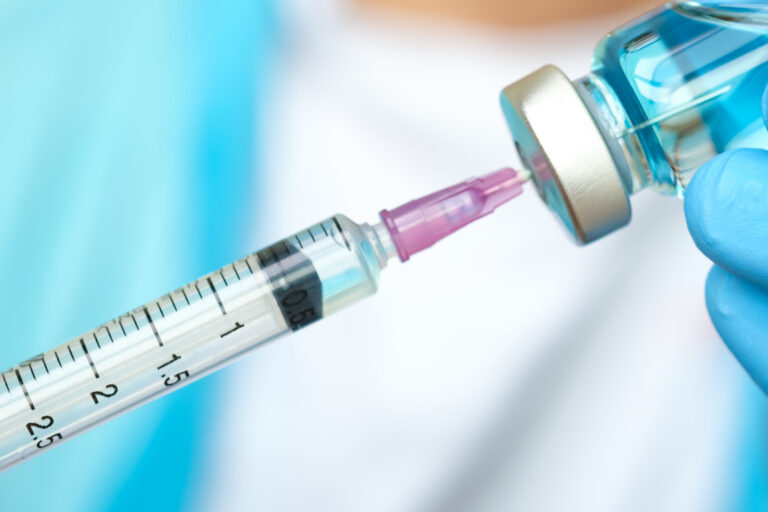
A great deal of research has focused on hepatitis viral disease in recent years. Joy Tomlinson, DVM, PhD, DACVIM, of Cornell University, described current information on this topic. To date, at least four different viruses with liver impacts have been identified. Two are considered of little consequence to horses because they live in the bone marrow and have little association with hepatitis: equine Pegivirus I and equine Pegivirus 2 (also called Theiler’s disease-associated virus or TDAV).
Equine Parvovirus-Hepatitis (EqPV-H) One of the two primary liver viral concerns is equine Parvovirus-Hepatitis (EqPV-H), which creates severe acute hepatic necrosis 4-12 weeks following administration of a biologic product such as tetanus antitoxin, plasma or allogenic stem cells. This causes a classic presentation of Theiler’s disease. However, EqPV-H has also been known to occur without use of biologic products with clusters occurring in summer and fall, indicating that it might be transmissible and contagious. The vast number of cases are mild or subclinical, and horses do not develop a fever. A prolonged period of viremia occurs prior to the onset of hepatitis. As the horse seroconverts and develops an immune response, the viremia level declines—at that point the horse experiences acute hepatitis. Tomlinson pointed out that viremia can persist at lower levels for months to years and that some horses do not completely clear the virus.
In the USA, 13–37% of healthy horses are PCR positive for EqPV-H, meaning that a single PCR positive test doesn’t confirm that a hepatic episode is caused by that virus. For accurate diagnosis, it is advantageous to combine a liver biopsy with a PCR test and to note the decline of viremia over time.
Typical transmission is due to iatrogenic biologic product administration, but recent USDA requirements test blood donor herds for biologics and allogenic stem cells. Tomlinson has identified that the virus is shed through nasal and fecal secretions for many weeks after infection, and because viral shedding starts prior to the onset of hepatitis, it is nearly impossible to protect others in advance. Nasal shedding occurs during a 6-19-week incubation. As yet, there is no evidence of vertical transmission, no vaccine, and no effective anti-viral drugs. By the time one sees disease, the horse’s immune system has likely already controlled the infection.
Equine Hepacivirus (EqHV) The second liver virus of concern is equine Hepacivirus (EqHV), a close relative of hepatitis C in humans. Hepacivirus tends to become a chronic problem in 20% of infected horses. Horses are more effective at controlling this virus, although they might experience prolonged viremia (for months). Subclinical hepatitis manifests at the time of viral clearance, similar to the pathophysiology of EqPV-H. A horse’s GGT and AST might increase significantly yet remain within normal limits. Affected horses are not overtly sick, and they have no fever although they develop acute hepatocytic necrosis and inflammation. Hepacivirus is cleared spontaneously by 80% of horses.
In the United States, 8-23% of horses have a positive PCR test for equine hepacivirus. Liver biopsy is not consistent for diagnosis. As viremia declines, it can take weeks to months to return liver enzymes back to normal. It is important to rule out other causes that need treatment, especially bacterial cholangiohepatitis, toxins or lipidosis.
Transmission of hepacivirus is through iatrogenic blood products, yet the USDA is not requiring testing at this time. It is unknown if mosquito and insect vectors are possible transmitters, but so far there is no evidence that is the case. There is some evidence of vertical transmission in utero. Like Parvo-Hepatitis, there is no vaccine nor is there treatment for hepacivirus. Both EqPV-H and EqHV are able to survive a freeze-thaw cycle.
Equine Coronavirus
SARS-CoV-2 aka COVID-19 No doubt, you’ve had clients ask you about potential acquisition of the COVID-19 virus in their horses. Nicola Pusterla, DVM, PhD, DACVIM, DAVDC-Eq, of the University of California, Davis, commented that equids are apparently susceptible to SARS-CoV-2 based on homology of the ACE-2 protein receptor. Experimental infection with MERS-CoV has not resulted in productive viral shedding or seroconversion, indicating that horses are not real susceptible. Of 667 nasal secretions submitted to UC Davis Diagnostic Lab, EHV-1 and 4, EIV and Strep equi were detected in 36%, but all were negative for SARS-CoV-2. Even in a horse that tested positive for SARS-CoV-2, the horse remained healthy with qPCR negative nasal secretions. Despite susceptibility, there was no apparent clinical disease of SARS-CoV-2 in that single individual.
In another study at the Del Mar racetrack, 22 asymptomatic jockeys and track workers tested positive for COVID-19. Serum taken from 63 Thoroughbreds ages 2-7 year identified a seropositive incidence in 6.5% of horses. However, the horses did not develop any clinical signs.
For a horse to be exposed, there needs to be contact with a shedding human. Forty percent of companion animals living with COVID-positive humans acquire infection and seroconvert. The biggest risk factor is if the companion animal sleeps with their humans. It was recommended that a human who is positive for COVID-19 take precautions with horses since there is always the potential for mutation that increases virulence in horses, which then could transfer back to humans.
Equine Coronavirus (ECoV) Equine coronavirus (ECoV) is a different story altogether, said Pusterla. As an enteric organism, infected horses do not always display enteric signs. Typical signs of ECoV include lethargy in 81%, anorexia in 91%, fever in 74%, watery diarrhea or soft feces in 21%, colic in 12% and encephalopathy in 4%. Neurologic signs develop due to hyperammonemia subsequent to necrotic enteritis.
This virus is transmitted via the fecal-oral route, mostly infecting adult horses. It has a morbidity of 10-83%. Sickness is usually self-limiting and is associated with low mortality. Case numbers are higher during colder months. Incubation takes 48-72 hours, and outbreaks last 2-3 weeks. Fecal qPCR is the preferred test over an antigen Elisa test. 90% of sick horses test positive for qPCR while 91% of subclinical cases test negative. Detection time is 3-9 days from onset of clinical disease, so the peak of viral shedding isn’t on the day the horse is tested. This provides plenty of opportunity for spread of the virus among a herd.
Potomac Horse Fever
Potomac horse fever, aka Neorickettsiosis, is an endemic seasonal infection of North and South America. Luis Arroyo, Lic Med. Vet, DVSC, PhD, at the University of Guelph, noted that this is the most frequent systemic entero-pathogen in diarrheic horses, yet it is challenging to confirm clinical disease. Most cases are mild, and 24% are subclinical. Neorickettsia risticii is a Gram negative, intracellular organism, making it difficult to culture.
Clinical signs are generalized – dullness (90%), anorexia (80%), intermittent fever (70%) ranging from 103-107 degrees Fahrenheit. Horses (70%) develop decreased gut sounds or ileus; congested and toxic mucous membranes; soft to profuse, watery, projectile diarrhea; mild colic (30%); dehydration; laminitis (20–25%); and mortality in 5-30%. It is also known to cause abortions at 90-150 days. This seasonal disease tends to occur in spring, summer, or fall with the highest incidence (in North America) in July and August.
The life cycle involves aquatic and terrestrial phases: Bats and adult birds eat insects and then develop flukes infected with the bacteria. Fluke eggs are then shed into water upon defecation where snails acquire the eggs and release cercaria containing the infectious agent. Then, cercaria are picked up by aquatic insects (mayflies, caddisflies), which hatch and enter creek, river or ditch water. Bats and adult birds eat the insects and the cycle continues. Disease is transmitted to horses that drink water or forage that is contaminated by aquatic insects. A horse becomes an aberrant dead-end host unable to spread disease. Environmental conditions affect numbers of intermediate hosts to impact prevalence of disease from year to year, and winds can blow flies for miles. It is recommended to turn lights off at the barn at night since lights attract flies that then die off and contaminate the area.
Nocardioform Placentitis
In 3-5% of Thoroughbred broodmares, placentitis is a problem that leads to late-term abortion. Pouya Dini, DVM, PhD, DECAR, DACT, of the University of California, Davis, described the four types of placentitis:
- Ascending placentitis, often due to B-hemolytic Strep equi and coliforms, that infect inward through the cervix;
- Diffuse placentitis, mostly from Leptospirosis, Salmonella,and Candidiasis;
- Multi-focal placentitis from bacteria and rarely fungi;
- Focal mucoid placentitis caused by Nocardioform bacteria.
Focal mucoid placentitis is found in the USA, South Africa, Italia, Australia and New Zealand. It most commonly affects the avillous area of the ventral placenta: 11% at the base of the uterine horns, 27% in the body; 26% in the non-gravid horn, 26% in the gravid horn and 4% at the cervical star. There is no involvement of allantoic fluid, amnion, amniotic fluid or the fetus.
Dini noted that an outbreak is usually preceded by hot and dry weather in August and September in the Northern Hemisphere. A broodmare experiences late-term abortion with weak yet viable foals or in some cases parturition is normal. An affected mare has normal fertility post-partum. Infection of the avillous area of the placenta interferes with fetal nutrition, but there is a negative correlation between lesion size and foal weight.
This infection is difficult to identify since a mare doesn’t commonly have a vulvar discharge unless she is about to abort. Transabdominal ultrasound is diagnostic to detect placental separation; however, if there is only a small lesion, it might not be visible. Treatment is based on that applied to an ascending placentitis—NSAIDs, progestin, trimethoprim-sulfa, doxycycline and/or gentamycin. These bacteria tend to be resistant to penicillin products. Treatment, however, Is not that successful, reported Dini. Seventy percent of pre-term, dysmature affected foals need treatment post-foaling.
Nocardioform is a gram-positive branching Actinomycetes. In a recent 2020 outbreak, only 54% of diagnostic testing identified bacteria; 45% were negative on culture and PCR.
The question Dini asked was, “Are other bacteria involved?”
Something might amplify the abundance of Actinobacteria while at the same time decreasing the number of Gram-negative Proteobacteria. This could then trigger inflammatory pathways. Dini said he suspected that dysbiosis was associated with an immune response due to an imbalance of microbiota in the placenta. The exact pathogenesis of this form of placentitis is still not known.
![[Aggregator] Downloaded image for imported item #19998](https://s3.amazonaws.com/wp-s3-equimanagement.com/wp-content/uploads/2026/05/29122748/EDCC-Unbranded-5-scaled-1-768x512.jpeg)