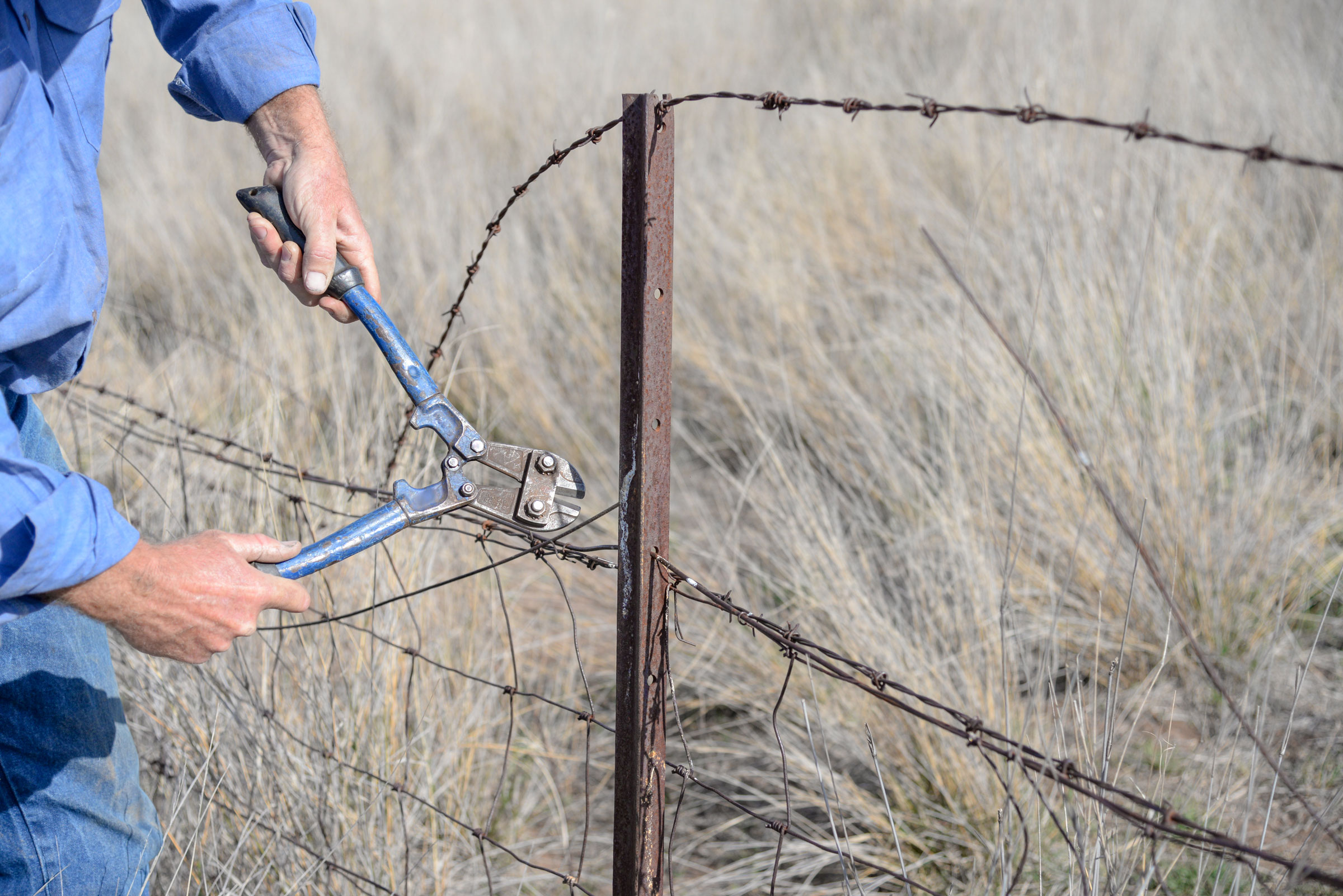
Although horses tend to be fairly circumspect about eating foreign objects, sometimes it is unavoidable. One dangerous culprit for the risk of colic is from ingestion of wire. In some instances, hay bales were wrapped with wire, although it is more common today to use baling twine. That said, there is always the possibility of wire or metallic foreign body ingestion, especially for inquisitive horses or for those eating out of tractor tire feeders (which might have steel belts that shred into feed). Also, needles—including acupuncture needles—are dropped accidentally; these and other wire pieces have been found in bedding.
Usual locations where wire foreign bodies are found are in the mouth, tongue, pharynx and cranial cervical region. A study sought to identify the incidence of wire in the equine abdomen as well as assess the diagnosis, treatment and outcome of such occurrences [Marley, L.K.; Soffer, C.; Hackett, E.S. Clinical features, diagnostic methods, treatments, and outcomes associated with ingested wires in the abdomen of horses: 16 cases (2002-2013). JAVMA Sept 15, 2018, vol. 253, no. 6, pp. 781-787].
A review of 11 years of medical records was undertaken for horses admitted to the Colorado State University Veterinary Teaching Hospital for colic, abdominal radiographs or exploratory laparotomy. In this retrospective study of 3,293 horses, wire was identified and confirmed in 16 horses via radiography or exploratory surgery. The primary presenting complaints included colic, lethargy and/or decreased appetite. Rectal palpation identified an abnormal abdominal mass in three of the horses.
Half of the horses had peritoneal fluid samples analyzed; all samples revealed median cell counts and protein that exceeded the upper limit of reference values. Bacteria were present in three of eight samples.
Transcutaneous ultrasound on all of the horses failed to identify the wire.
Of the three horses with abnormal abdominal masses, transrectal ultrasonography was performed on two; wire was only detected in one of those.
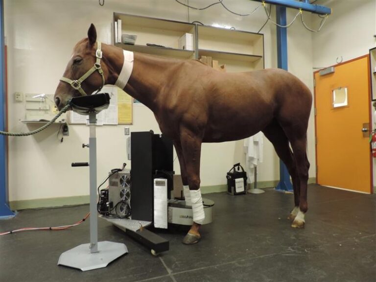
Abdominal radiography of seven of the horses identified a single wire in four horses and multiple wires in two horses.
The authors noted that of 149 cases of abdominal radiography at the teaching hospital, six had wire, which was sometimes an incidental finding.
Medical treatment (antibiotics, IV fluids) alone was administered to six of the 16 horses. All six were euthanized due to a progression of clinical signs, especially since treatment was unable to address the underlying cause. One of the 16 horses in the study was dead on arrival at the hospital.
Eight of the 16 horses underwent exploratory laparotomy in conjunction with medical treatment. Four of these were euthanized due to adhesions, abscesses or intestinal perforation resulting in abdominal contamination and peritonitis. Three horses had small intestinal perforation and the other had a large colon sand impaction containing a wire.
Of the 16 horses in the study, only four survived, recovered and were discharged. Those four were managed with both surgery and medical treatment. Necropsies on the non-survivors identified wire within sand or abscesses in the large colon, spleen, liver, diaphragm or thorax. The length of the wire foreign bodies ranged from 4-15 cm. Fourteen horses had wire penetration of the GI tract, making tetanus prophylaxis also important when dealing with ingested metallic foreign bodies.
In summary, the overall case fatality rate was 75%. For those treated successfully with surgery and medical intervention, the long-term survival rate was excellent.
While transcutaneous abdominal ultrasound does not necessarily identify a metallic foreign body, it is useful for diagnosis of splenomegaly, abdominal effusion, intestinal dilation and soft tissues masses; many of those are associated with ingested wire. The authors urged the use of abdominal radiography for horses with peritonitis or abdominal abscesses in order to enable the definitive treatment with surgery in a timely manner upon identification of a metallic foreign body.