An equine practitioner might be involved with horses through his or her entire career without ever encountering a hoof keratoma. However, it is a possible contributor to lameness and should be included in a list of differential diagnoses for foot pain.
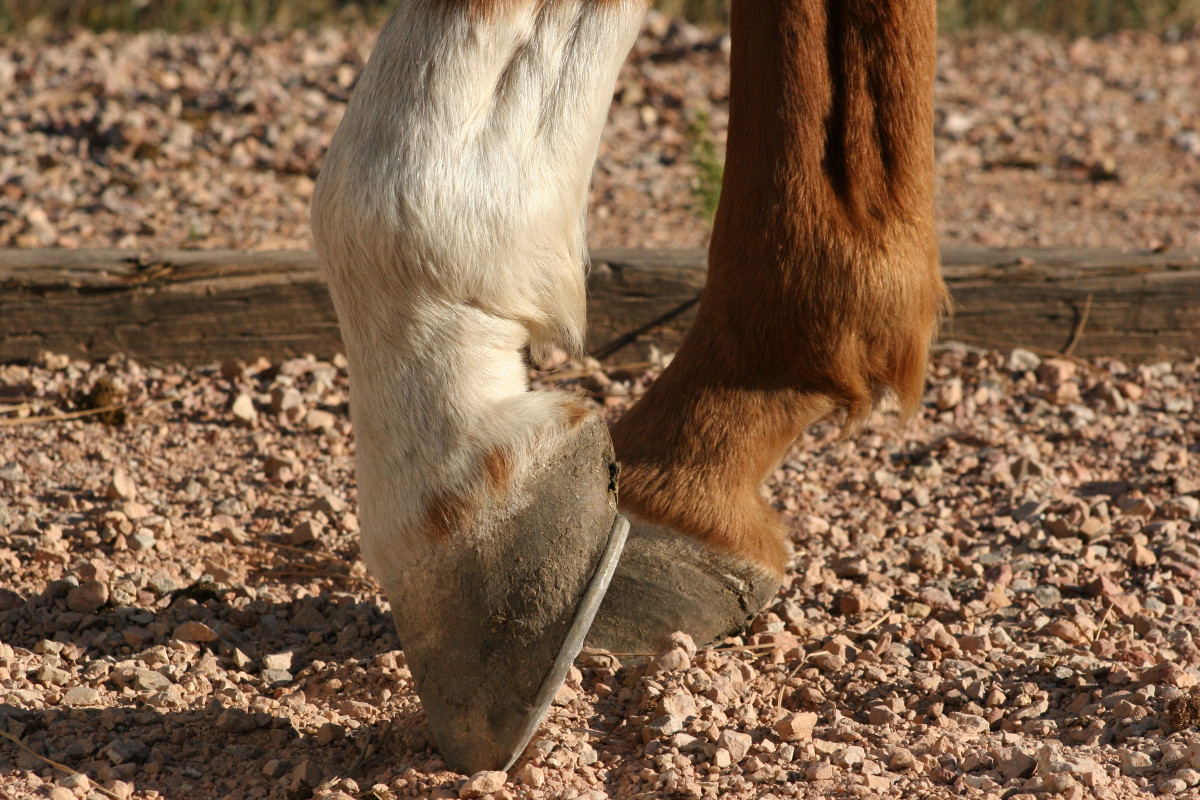
A keratoma results from the abnormal proliferation of keratin and squamous epithelial cells to form a benign (non-neoplastic), space-occupying mass within the hoof corium between the hoof wall and the coffin bone. Size normally ranges from a few millimeters up to 2-3 centimeters. Pressure from the invasive mass causes pain and progressive lameness as the lesion grows.
The inciting cause is not completely understood. It is thought that chronic irritation to the coronary band, the dorsal laminae or the solar corium might occur “due to injury, pressure, abscess formation, or chronic pododermatitis” that leads to scar tissue and keratoma formation. Local surgery might be another inciting cause.
There is no age or breed predisposition, although this study included a higher proportion of Warmblood breeds consistent with the population of horses in the study area.
These are usually solitary lesions and usually start growing just distal to the coronary band. The hoof is often deformed, particularly on the dorsal or lateral wall; the white line might also deviate. Sufficient disruption of hoof architecture can lead to abscess around the lesion.
A retrospective study [Joana Sofia Terra e Pereira. Retrospective Study of Keratoma Lesions. Universidade de Tras-os-Montes e Alto Douro, 2017] reviewed 21 cases over an eight-year period at the Gent University Equine Hospital in Belgium. Thirty-eight percent the horses had a history of hoof abscesses; 29% had a history of hoof cracks; 5% had white line disease. Thirteen of the 21 horses (62%) were lame; the others (38%) were not.
While it is often difficult to definitely diagnose the condition with radiography, 87% had diagnostic radiographic findings, some with osteitis or radiolucency within the coffin bone. Following hoof resection, half the horses developed abnormal hoof configurations. Eight percent became sound with recovery corresponding to the time it took for hoof regrowth. Within 1-4 years following keratoma removal, 60% experienced keratoma recurrence, likely due to incomplete removal of the keratoma tissue at surgery. Other studies report only a 30% recurrence rate.