
Editor’s Note: Vetscan Imagyst™ gives veterinarians a straight line to a clinical pathologist available 24 hours a day who can quickly provide interpretation of cytology samples.
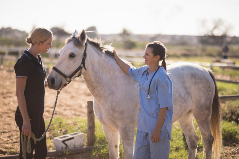
Laura H. Javsicas, VMD, DACVIM, is a veterinarian at Rhinebeck Equine in New York. Janine Baker, LVT, is a licensed veterinarian technician and lab manager at that clinic working with Javsicas in internal medicine. We spoke with this team to find out how they are using Vetscan Imagyst, a new multispecies diagnostic platform from Zoetis. Rhinebeck uses the digital cytology tool and is an early experience tester of the equine fecal egg count system on Vetscan Imagyst. What we discovered is that Vetscan Imagyst was beneficial not only to equine veterinarians, but also to techs, horses and owners.
What is Vetscan Imagyst
Vetscan Imagyst is a multi-use platform that allows board-certified clinical pathologists to remotely examine cytology slides and uses artificial intelligence (AI) to evaluate fecal samples digitally. As new applications become available, they will be easily integrated with existing Vetscan Imagyst testing capabilities.
For cytology, the practice can easily create slides from any specimen type and send images of those slides to a board-certified clinical pathologist. Clinical pathologists are available 24 hours a day. Pathology reports are returned to the veterinary practice within two hours as compared to a traditional cytological evaluation, which can take 24 hours or longer for a final report after slides are sent out.
Artificial intelligence (AI) fecal egg counts for small animals is currently available, with AI equine fecal egg counts becoming available in the Spring of 2023.

When and Why Did You Start Using Vetscan Imagyst?
Javsicas said she had been looking for a “tele-cytology” solution for the practice before the 2021 AAEP Convention. “I liked the idea of the rapid turnaround with Imagyst,” she said. Through Vetscan Imagyst, clinical pathologists are available to give interpretations of the cytology, and fecal slides are analyzed with AI and are available for consultation.
After talking with Zoetis reps at the 2021 AAEP Convention, Vetscan Imagyst was purchased and installed at Rhinebeck Equine in January of 2022.
“Our Zoetis rep came out and set up the equipment and walked us through using it,” said Baker.
Baker said there was a “learning curve” to using the new equipment. Once she understood what steps needed to happen, she taught the practice’s other techs. There is a flow chart of steps provided by Vetscan Imagyst. There also are videos available online for refresher training on the different applications. Baker said training all the techs in the practice allows the equipment to be used 24/7/365 no matter who is on duty.
“It’s user-friendly once we used it a bit,” said Baker. “But the [clinical] pathologist can only give a good report if the slides are properly prepared. That takes some skill.”
How Vetscan Imagyst Works for Rhinebeck
The Vetscan Imagyst digital whole slide scanner does not take up much room. You also need a small space to prepare samples. The equipment is integrated with your practice management software, and you need a direct connection to high-speed internet for best results.
The cytology sample is prepared according to instructions and a slide is created, stained and dried. A 24×60 mm coverslip is then applied to the slide with immersion oil and the slides are inserted into the Vetscan Imagyst scanner. The images from those slides are reviewed by the tech, with areas selected for pathologist review. Then the images are submitted via a computer form (that includes case identification) to the pathologists on duty.
“You can follow in real time on the web where your sample is in the process,” said Baker. “And there is always a [clinical] pathologist available to review. They send the report, and you have access right away.”
Types of Cases for Vetscan Imagyst Use
Javsicas said there are multiple cytology uses for the Vetscan Imagyst equipment and services, including peritoneal fluid, joint fluid or fine-needle aspirates.
“We just had a great ophthalmology case” where the Vetscan Imagyst equipment helped immensely. A horse previously had a third eyelid hemangiosarcoma mass removed. The horse came back, and the veterinarian suspected the hemangiosarcoma had recurred even though clean margins were obtained with the first surgery.
“Our ophthalmologist who comes to the clinic once a month,” said Javsicas. “She was there that day, so we took the cytology sample, and the pathology report was consistent with recurrence of hemangiosarcoma cancer. I performed abdominal radiographs and a rectal exam check for metastasis. Because of the quick answer, the ophthalmologist was able to remove the eye that day, decreasing the chance that the tumor would spread.”
Baker said, “I have sent out (samples) in the day, evening and weekends … It’s always a fast report, within two hours, and usually faster!”
Javsicas added, “It is a new test, so we need to remember what it can do. With select cases, it is really helpful to get results quickly (within two hours). [When sending in physical slides for evaluation, traditionally] it’s at least 24 hours. This can help answer the questions such as ‘does the horse need surgery?’ or ‘should we lavage the joint with arthroscopy with the horse under anesthesia or can we just flush?’
“It certainly can impact the quality of care,” she stressed.

Client Feedback
Javsicas said her practice runs labs for referring veterinarians. “There is not a lot of demand now, but they are learning (about Vetscan Imagyst),” she said.
For owners, she cited the eye case mentioned above. “The owner appreciated we could get quick results and do the surgery rather than the horse go home and have to come back at a later date,” Javsicas said.
Baker added that “this is another tool in our diagnostic toolbelt. And we’re becoming more willing to reach for it because of the speed [of access to a final report]. If you know you have to wait 24 hours or more for a cytology [report using traditional methods of sending slides in], you might not do it.”
She added that “owners happily pay for the time and expertise.”
Sometimes test results are negative. Such as peritoneal fluid that has no neoplastic cells. Baker said clients are happy to have the results at the time of examination for quicker peace of mind. “I looked back at some of our cases, and a lot of the results are ‘not bad.’ But that’s a benefit!” Baker added.
Benefits for Practices and Technicians
Equine veterinary practices are looking to better utilize the skills of their technicians and assistants. “This technology empowers equine techs and allows them to utilize new skills,” Baker added.
“This technology really empowers techs!” Baker added.
Working with the Clinical Pathologists
Baker said you can select on the slide images the areas you want scanned by the clinical pathologist. With a blood smear, she said you want a mono-layer and a feathered edge. “It’s harder with peritoneal fluid,” she noted. “You can submit two slides per peritoneal sample.”
She said part of the learning curve was making the slides and staining them, then confirming and adjusting the recommended scan areas for the clinical pathologist to review.
“The pathologists are good at giving tips,” said Baker. “And they are always available for consultation. They even compliment us on good slides and like to follow up on cases.”
Fecal Egg Count Ability
When Javsicas was first talking to Zoetis about Vetscan Imagyst, she was primarily interested in tele-cytology option horses.
For small or mixed animal practices, there is the option for digital AI (artificial intelligence) to read a fecal slide for a dog or cat in minutes (while the client is still at the office). Additionally, there is the ability to flag a result and request a consult with a boarded specialist, if needed.
Without Vetscan Imagyst, fecal sample results are read in-house by technicians—which can be time-consuming and relies on the knowledge and experience of the person reading the slide.
However, the Vetscan Imagyst developers have focused on improving the AI equine fecal egg count options for horses, which can take the human error factor out of the equation.
While there are other automated fecal analyzer options for horses, most are not able to differentiate equine parasite eggs and have a lot of required supplies. “We see a lot of foals, so we need to differentiate between ascarids versus strongyles,” said Javsicas.
She said Rhinebeck has worked with Zoetis to fine-tune the equine fecal testing resources available today. Those resources include a report that is “clean and client-friendly,” said both Javsicas and Baker. The report includes images of the parasite eggs counted.
Baker said Rhinebeck does fecal egg counts in-house, counting the eggs by hand. “We do them manually in-house, and it takes a lot of time,” she added.
Rhinebeck was an early experience tester of the equine fecal egg count system on Vetscan Imagyst, Baker said. “We are doing all the manual counting and compare the results to the Vetscan Imagyst,” she said. “The results have matched well,” she stated. In those rare instances in which there is a discrepancy, Baker said that they “can figure out why we are getting different results.”
She noted that, “The report is very client-friendly and includes images of the eggs counted.”
Zoetis stresses that use of the Vetscan Imagyst AI fecal egg count can improve a practice’s accuracy of results, speed of results and provide improvement in overall efficiency for technicians. This application will be universally available in the Spring of 2023.
From Zoetis
“Every veterinarian knows the importance of easy and rapid access to specialists when diagnosing and treating our patients,” said Richard E. Goldstein, DVM, DACVIM (SAIM), DECVIM-CA, Vice President and Chief Medical Officer, Global Diagnostics at Zoetis. “With the new digital cytology solution available from Vetscan Imagyst, we are virtually placing a board-certified, clinical pathologist right ‘down the hall’ from every veterinarian.”
The Vetscan Imagyst Resources page (https://www.vetscanimagyst.com/resources) includes downloadable papers and training videos for companion animals. The slide preparation videos are simple to follow and are pertinent to any species. Equine-specific resources will be available in the Spring of 2023.
About Dr. Laura Javsicas
Laura Javsicas, VMD, DACVIM, received a B.A. in Biology from Cornell University, where she was co-captain of the equestrian team. She then attended veterinary school at the University of Pennsylvania. After receiving her VMD in 2004, she did an internship at the Hagyard Equine Medical Institute in Lexington, Kentucky. Javsicas then completed a three-year residency in equine internal medicine at the University of Florida. Following her residency, she stayed at the University of Florida as a member of the faculty. In 2009, she moved to Saratoga Springs, where she worked at an equine clinic providing in-hospital internal medicine services and general ambulatory care until joining Rhinebeck Equine in 2013. She has special interests in neonatology, ultrasonography, cardiology, gastrointestinal disease and emergency/critical care medicine. Javsicas has served on Cornell’s Zweig Committee since 2016.
About Janine Baker, LVT
Janine has a B.T. in Equine Science from SUNY Morrisville and a Veterinary Technician A.S. from Penn Foster College. Janine has worked at Rhinebeck Equine since 2015 as the internal medicine technician and laboratory manager.
About Rhinebeck Equine
Rhinebeck Equine is an independently owned, exclusively equine practice located in Dutchess County in the Hudson River Valley about 120 miles north of New York City. The practice’s veterinarians have been providing quality care to the equine population of New York’s Hudson Valley for over 50 years. This experienced and trusted practice has progressively grown into a comprehensive ambulatory and referral clinic with 12 veterinarians, five intern veterinarians and more than 30 support staff.
The Rhinebeck Equine LLP team offers a full spectrum of services to horses of all breeds, providing comprehensive equine health care, including services in the areas of surgery, sport horse care, lameness evaluations, pre-purchase exams, reproduction, neonatal care, internal medicine, ophthalmology, dentistry and wellness care.
Rhinebeck Equine’s referral hospital is staffed by board-certified specialists in equine surgery, internal medicine, theriogenology (reproduction), ophthalmology, dermatology, interns and a team of highly experienced support personnel. The hospital enables Rhinebeck Equine LLP to provide cutting-edge treatment to its equine patients. Ambulatory veterinarians serving the surrounding area provide care from their fully equipped mobile units.


![[Aggregator] Downloaded image for imported item #19998](https://s3.amazonaws.com/wp-s3-equimanagement.com/wp-content/uploads/2026/05/29122748/EDCC-Unbranded-5-scaled-1-768x512.jpeg)