Imaging of the distal limb is fundamental for the diagnosis and treatment of problems with the foot and fetlock. The adage no foot no horse remains as true today as it was back in 1895, when the equine foot was one of the first veterinary radiographs to be published. The publications Veterinary Radiology & Ultrasound (VRU), Equine Veterinary Education (EVE) and the Equine Veterinary Journal (EVJ) have combined forces to celebrate the evolution in equine imaging with a free special collection to reflect some of the most significant advances in distal limb imaging from the past five years.
Advances in Imaging of the Equine Distal Limb 2017–2022 comprises a total of 20 papers which have been selected by Mathieu Spriet, Ann Carstens and Tim Mair. It is accompanied by a comprehensive editorial from the EVJ summarising the major historical technological developments in imaging of the foot and fetlock, embracing all the modalities.
The evolution of computed tomography (CT) allows the imaging of the distal limb without anaesthesia. CT scanners are also now used in surgery rooms bringing significant progress in orthopaedic surgery. Six papers look at advances in CT, including addressing some of the challenges of the ring design of this modality.
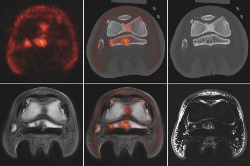
Positron emission tomography (PET), the latest modality to be introduced to equine imaging, has opened a whole new field of possibilities for bone and soft tissue imaging in racehorses and sport horses. Five papers look at how PET brings functional information to the table, allowing early detection of abnormalities before the occurrence of structural changes and distinguishing between active and inactive lesions when structural changes are present.
MRI has been a mainstay in orthopaedic imaging for many years. Six important studies are included in the collection and show how the optimization of scanning techniques is constantly improving this modality.
Ultrasound is steadily improving. One of the papers looks at the important technical evolution involving the imaging of limbs in non-weight bearing position as well as under the classic weight bearing position.
Radiographs remain the most commonly used imaging technique, despite the modality being more than 100 years old. One of the papers confirms why radiographs remain a valuable imaging tool.
The role of scintigraphy has decreased in the past 20 years with the emergence of advanced cross-sectional imaging, but it remains an essential tool especially for comparative imaging studies. One of the papers examines agreement between scintigraphy and MRI to identify the source of foot pain.
“The content of this virtual issue represents an amazing amount of new knowledge that with no doubt will contribute to improve equine welfare and safety,” said Mathieu Spriet. “With the increase availability and versatility of all the imaging modalities, the knowledge base appears to increase exponentially. We are very excited to see what the next five years will bring. We hope the readers will enjoy consulting this collection as much as we enjoyed putting it together.”
“During the past five years close to 100 equine imaging papers have been published between EVJ, VRU and EVE, covering a wide variety of topics,” said Professor Celia Marr, Editor of the EVJ. “The EVJ is proud to have been able to work collaboratively to bring this definitive distal limb imaging collection to life, giving clinicians easy access to some of the most pertinent work in this area.”
The virtual issue is free for 12 weeks and can be found at https://beva.onlinelibrary.wiley.com/doi/toc/10.1001/(ISSN)2042-3306.equine-distal-limb
![[Aggregator] Downloaded image for imported item #19769](https://s3.amazonaws.com/wp-s3-equimanagement.com/wp-content/uploads/2026/05/31141823/EDCC-Unbranded-11-scaled-1-768x512.jpeg)

