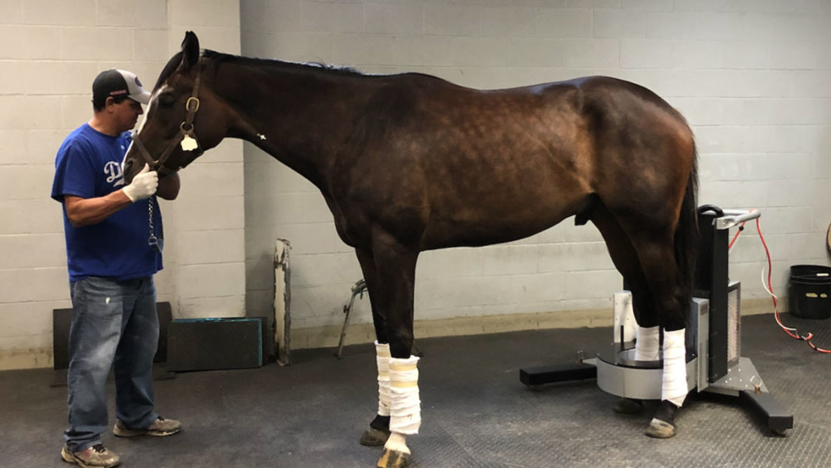
On December 12, 2019, Santa Anita Park Installed the world’s first MILE-PET device, a positron emission tomography (PET) scanner specifically designed to image standing racehorses. This installation, one of several measures to reduce breakdowns at the racetrack, received a lot of attention at a time when Santa Anita was just coming out of a challenging racing season, with a cluster of horse fatalities early in the year.
The Stronach Group, owner of Santa Anita, invested in equine PET following the promising pilot work at the UC Davis School of Veterinary Medicine. Over the previous four years, the UC Davis team demonstrated that PET could detect abnormal changes in bones of racehorses that were not seen with other imaging modalities. This initial work, however, required horses to be laying down under general anesthesia, a major barrier to common use at a racetrack.
Inspired by the success of the early studies, LONGMILE Veterinary Imaging developed the MILE-PET scanner for safe imaging of standing, lightly sedated horses.
Where do things stand a year later?
“Things went really well!” said Mathieu Spriet, DVM, DACVR, DECVDI (Diplomate American Collage of Veterinary Radiology and Diplomate European College of Veterinary Diagnostic Imaging), a veterinary radiologist at UC Davis who leads the research on equine PET. “In the past 12 months, 188 PET studies were successfully performed at Santa Anita on more than 100 different horses, with many horses imaged multiple times to follow the evolution of bone changes over time.”
The MILE-PET scanner performed robustly, without a single down day. About half of the PET scans were part of sponsored research projects, while the other half were requested by racetrack veterinarians for specific assessment of horses under their care.
One of the research projects, sponsored by Grayson Jockey Club Research Foundation, allowed comparing findings of PET scan with those of nuclear scintigraphy (bone scan), an imaging technology that was used at Santa Anita for more than 20 years. The fetlocks (ankles) of 32 horses were imaged with both modalities and the data were independently analyzed by a group of seven different observers, including racetrack veterinarians and UC Davis researchers. The results showed that veterinarians were able to more consistently detect and better characterize abnormalities on PET scans than on bone scans. This was particularly true for abnormalities involving the sesamoid bones—small bones at the back of the ankles that are commonly involved in catastrophic fetlock fractures.
In a second part of the Grayson Jockey Club Research Foundation study, 24 horses were followed over time, with scans repeated at 6 and 12 weeks after the initial scan. This project determined how quickly abnormalities might improve over time in horses resting from training. Preliminary results demonstrated that some abnormalities respond positively to rest quickly, whereas others are more problematic and require a longer time for healing.
Another project recently funded by the Dolly Green Research Foundation focuses on how bone responds to training after rehabilitating from an injury. Five sequential PET scans will be performed over a 6-month period to monitor horses reentering training after recovering from injury. A total of 12 horses will be entered in this study; seven have already been enrolled.
“We have learned a lot through these different projects,” said Spriet. “We recognize a few common patterns, but each horse is different.”
Some of the findings are considered normal adaptation to training, but some of the changes occurring in specific areas might predispose a horse to severe injury. Each PET scan is evaluated in light of knowledge of the characteristics of fractures observed through the California Horse Racing Board post-mortem program that documents the configurations of fatal injuries sustained on racetracks. A specific spot in one of the sesamoid bones has been identified as being commonly abnormal in horses with fetlock breakdowns. When the PET scan shows abnormalities in this area, horses are considered at higher risk for fractures.
The results of PET scans have contributed to many decisions regarding horses’ careers. In several cases, the PET findings have contributed to the decision to retire a horse. In other horses, the abnormalities have been followed over time to monitor healing and identify the right time to resume training. Several of these horses are now racing successfully.
In combination with other measures to prevent fatalities, such as the use of MRI scans, pre-race veterinary inspections and changes in medication rules, PET has contributed to a safer racing season at Santa Anita. No racing fatalities occurred during the fall meet.
PET has established itself as a reliable and helpful diagnostic tool at the racetrack, but there is still a lot more to learn. An upcoming project will use PET to look at horses performing well, thus helping to distinguish between normal adaptation to training and developing injury.
PET will also be used to scan horses with repaired fractures to assess existing injuries that might have led to the fractures and to monitor bone healing.
PET innovation and success at Santa Anita inspired the installation of a standing PET scanner on the East Coast, at the New Bolton Center at the University of Pennsylvania.
With support from the UC Davis Center for Equine Health and the Stronach Group, a MILE-PET scanner will be installed at the UC Davis veterinary hospital in early 2021, making this technology available for racehorses in Northern California.
The availability of these scanners at multiple sites will enable comparing injuries on different racetracks, improving understanding of possible causes for injuries, and helping further prevention of breakdowns.
This article was reprinted from the UC Davis School of Veterinary Medicine and was authored by Rob Warren.