In this episode of the Disease Du Jour podcast, Amy Johnson, DVM, DACVIM (LAIM & Neurology), discussed noninfectious neurologic diseases in horses, specifically cervical vertebral stenotic myelopathy (CVSM) and equine neuroaxonal dystrophy (eNAD)/equine degenerative myeloencephalopathy (EDM). She described clinical signs, diagnostics, management options, and more.
Clinical Signs of Noninfectious Neurologic Disease
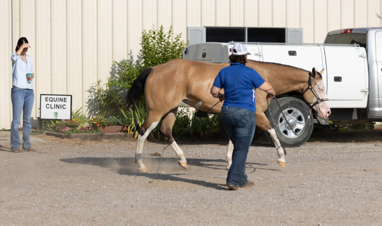
Johnson said performance decline is a common early sign of neurologic disease for horses in work. The horse might knock rails, refuse jumps, trip under saddle, display a toe drag, or struggle to move up the levels in dressage.
While infectious neurologic diseases have a rapid onset and progression (EPM is one exception), noninfectious diseases usually progress slowly. Johnson said symmetric muscle atrophy is more indicative of noninfectious disease, whereas asymmetry is more common with infectious disease. Most horses with infectious disease will also present with a fever.
CVSM in Horses
Johnson explained that CVSM is caused by spinal cord compression due to malformation or abnormal development of the vertebral column. It is especially common in young, rapidly growing horses like Thoroughbreds, but she also sees the condition in warmbloods and older horses.
“We know that spinal cord compression can happen because of malformation of the bones of the neck itself, sometimes because of the onset of degenerative joint disease or arthritis in the neck that leads to a slower spinal cord compression,” Johnson said.
Diagnosing CVSM typically starts with survey cervical radiographs. “You can’t diagnose the presence of spinal cord compression on survey radiographs, of course, because you can’t visualize the spinal cord, but there are certain morphologic features in the bones that might make CVSM more or less likely if you see them,” Johnson explained. Findings that could raise suspicion of CVSM include evidence of narrowing of the vertebral canal, clear osteochondral fragments or degenerative joint disease of the articular process joints, or malalignment of the vertebra.
Whenever possible, Johnson recommends advanced imaging to confirm a diagnosis. Currently, the best imaging option available is CT myelography. “CT is great because it provides cross-sectional imaging, and so unlike the standard survey myelogram that gives us a lot of information about dorsoventral compression, the CT allows us to see lateral and dorsolateral compression that previously we probably missed,” she explained.
Treatment options for CVSM include conservative management and surgery. A conservative approach might involve reducing the horse’s workload and giving anti-inflammatories either systemically or intra-articularly to decrease inflammation surrounding the spinal cord. “In some cases, say in the horse that has a stenotic canal but not spinal cord compression all the time, the conservative management might be enough,” said Johnson.
If compression is consistent, Johnson said, conservative management will probably eventually fail. “In that case, the most common surgical procedure performed is cervical fusion surgery, whereby two adjacent vertebrae are fused with the traditional basket implant or with some of the newer implants that involve spacers and little plates,” she said.
Johnson said up to 80% of horses that undergo surgery will improve, and 50-60% will return to their intended purpose. Conservative management has a lower success rate; one study looking at young Thoroughbreds being managed conservatively reported that about 30% returned to racing.
eNAD/EDM in Horses
eNAD and EDM are neurodegenerative diseases that “may very well be a spectrum of the same neurodegenerative disorder,” said Johnson. eNAD/EDM has been described in many different breeds, including Quarter Horses, Arabians, Morgans, Thoroughbreds, and Standardbreds. Johnson’s case population includes many young to middle-aged warmbloods.
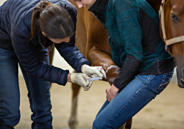
Neurodegenerative diseases can be tricky to diagnose. “In the living horse, they are a diagnosis of exclusion, meaning we don’t have any tests that will tell you definitively that the horse has the disease,” said Johnson. That process often involves imaging to rule out CVSM and spinal fluid analysis to rule out EPM. Johnson might also biopsy the horse’s tailhead muscle to check for evidence of past vitamin E deficiency. However, the only definitive diagnostic procedure is postmortem evaluation of the brain and spinal cord.
Neurodegenerative diseases are progressive, which makes them challenging to manage. Johnson said horses often benefit from ridden or ground exercise, if it’s safe to do so. “The more the horse exercises, often the better they do, because it maintains their muscle strength, it maintains coordination, and it allows them to develop compensatory pathways for neurons that have been lost,” she said. Vitamin E supplementation is also helpful.
Prognosis for eNAD/EDM is variable. Johnson has encountered horses that deteriorate rapidly within a year and necessitate euthanasia based on the severity of their ataxia or behavior changes. She’s seen other horses that have remained in work for years. “The owner’s commitment to the horse as well as their willingness to accept risk does play into it a little bit,” Johnson said. In general, horses that develop mild ataxia early in life and then stabilize tend to do better than horses that develop ataxia later in life or demonstrate substantial behavior changes.
Final Thoughts
In closing, Johnson encouraged veterinarians to keep an open mind if they encounter a horse displaying odd behaviors, performance problems, or a tricky lameness. “Remember the possibility of neurologic disease, because they can sometimes occur very insidiously and slowly over time,” she said. “The sooner you recognize that neurologic disease might be one of the contributing factors to the horse’s problems, the sooner you can address it, and hopefully that will result in a better prognosis for the horse.”
About Dr. Amy Johnson
Amy Johnson, DVM, DACVIM (LAIM & Neurology), is board-certified in large animal internal medicine and neurology and was the first American veterinarian to obtain these credentials. Her clinical caseload consists predominantly of horses with neurologic problems, including spinal cord compression, neurodegenerative disease, and infectious diseases. She also provides consultations for farm animals with neurologic disease. Her research efforts focus on improving our ability to diagnose the cause of neurologic disease in horses. Johnson has worked at PennVet’s New Bolton Center since 2007 and is the current Section Chief of Internal Medicine and Ophthalmology.
Related Reading
- Differentiating Noninfectious Neurologic Diseases in Horses
- Vitamin E Response for eNAD and EDM
- Ventral Interbody Fusion for Horses With C7-T1 Pathology
Stay in the know! Sign up for EquiManagement’s FREE weekly newsletters to get the latest equine research, disease alerts, and vet practice updates delivered straight to your inbox.