In this episode of the Disease Du Jour podcast, Kelly Knickelbein, VMD, DACVO, joined us to discuss ocular squamous cell carcinoma (SCC) in horses, including clinical signs, risk factors, treatment options, prevention strategies, and more.
This episode of Disease Du Jour is brought to you by Equithrive.
What Is Ocular Squamous Cell Carcinoma?
Ocular squamous cell carcinoma is the most common tumor to affect the horse’s eye. It is an epithelial tumor that typically develops on the eyelids, third eyelid, and ocular surface, including the conjunctiva and superficial cornea. When tumors arise on the ocular surface, they are usually located on the lateral bulbar conjunctiva near the lateral limbus, although they may appear anywhere on the conjunctiva or limbus and often extend onto or within the cornea.
Knickelbein explained that SCC develops when ultraviolet (UV) exposure causes DNA damage. When cells cannot adequately repair this injury, normal cell-cycle regulation fails, and uncontrolled cellular proliferation can result in a tumor.
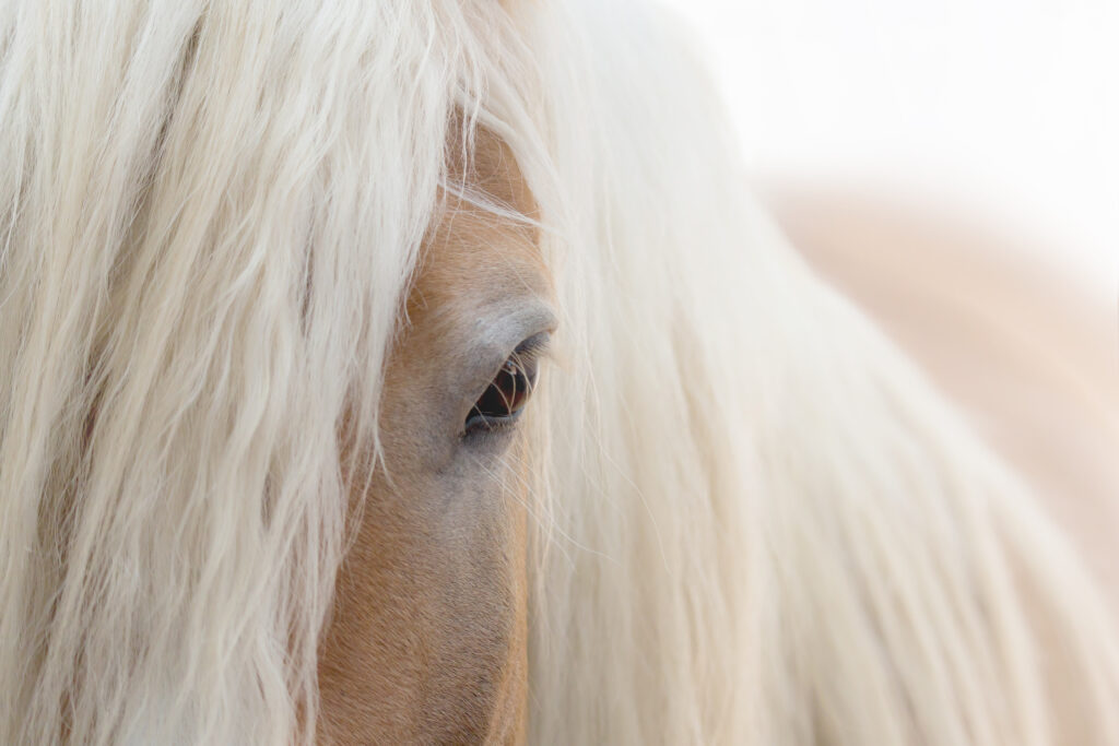
What Types of Horses Are Most at Risk?
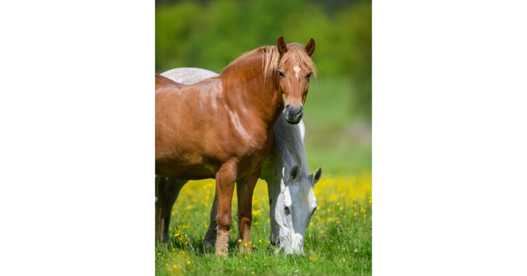
UV exposure is a primary risk factor. Horses with reduced periocular pigmentation, particularly those lacking melanin in the eyelids and conjunctiva, are at increased risk. Breeds frequently affected include Paints, Quarter Horses with extensive white facial markings, and Appaloosas.
Genetics are another known risk factor. A research team led by Rebecca Bellone, PhD, at the UC Davis Veterinary Genetics Lab identified a missense mutation in the gene DDB2, which is involved in UV-induced DNA damage detection and repair. Horses with two copies of the variant are at significantly higher risk of developing ocular SCC. The mutation was first identified in Haflingers but has since been reported in Belgian Drafts, Rocky Mountain Horses, Connemaras, Holsteiners, and Belgian Warmbloods.
“This genetic variant changes the structure of the protein so that it can no longer scan the genome to find UV damage,” Knickelbein explained. “Thus, the damage goes unrepaired and tumors result.” The variant is inherited in a recessive pattern; current evidence does not support an increased risk of ocular SCC in heterozygous horses.
Clinical Signs of Ocular SCC
Clinical signs vary based on lesion location and stage. The first sign of third-eyelid or ocular surface SCC is often a thick white or yellow ocular discharge. “Most of the conjunctiva is hidden underneath the eyelids, so a tumor can be growing that’s not directly visible but is producing this discharge,” Knickelbein explained. “Any horse that has unexplained thick ocular discharge should have a really thorough visual examination of all of the conjunctival surfaces as well as digital palpation of these surfaces and behind the third eyelid to see if there is a tumor hiding.”
When visible, early conjunctival or third-eyelid lesions may start as flat white or pink plaques that progress to pink, raised, proliferative masses. Eyelid SCC usually presents along the eyelid margin as hyperemia, erosions, or ulcerations.
Habronemiasis (summer sores) can resemble SCC. These lesions often contain yellow granules and occur at the medial canthus. “It can be hard to distinguish the two clinically, so in these cases, biopsy is your best bet to ensure you know what it is that you’re dealing with,” Knickelbein said.
SCC is invasive and can spread into the local tissue surrounding the primary tumor site. Eyelid tumors can invade through the skin to the underlying muscle and bone. Third-eyelid tumors can invade the orbit and wrap behind the globe. Lesions in the medial canthal region can invade the nasolacrimal duct, providing direct access to the skull.
Treatment Options for Ocular SCC
Early diagnosis and intervention are critical. “This truly is a management disease,” said Knickelbein. Even after successful treatment of one lesion, affected horses can develop SCC at additional sites, often requiring ongoing monitoring and repeated treatments.
Treatment typically involves surgical excision of as much of the tumor as is feasible. Because achieving complete tumor-free margins through surgery alone is difficult, surgery is typically combined with adjunctive procedures. (Exceptions include tumors confined to the leading edge of the third eyelid, which can often be cured with complete third-eyelid excision.)
Adjunctive options include intralesional or topical chemotherapy, radiation, cryotherapy, carbon dioxide laser therapy, and photodynamic therapy.
What Is the Prognosis for Ocular SCC?
Prognosis depends heavily on lesion size and location and the timeliness of treatment. Small tumors on the leading edge of the third eyelid that are fully excised with clean margins have a good prognosis. Early ocular surface SCC also has a favorable outcome when treated promptly with surgical excision and adjunctive therapy.
Advanced disease carries a poorer prognosis, especially when bone involvement is present. Extensive third-eyelid SCC can invade the orbit and nasolacrimal system, making complete excision unlikely. Once bone is affected, management becomes palliative, and humane euthanasia is often warranted due to pain.
Horses with genetic predisposition appear to develop SCC at a younger age, increasing the lifetime likelihood of recurrence or new tumor development. However, there is currently no evidence that the DDB2 variant results in more aggressive tumor behavior.
Prevention Strategies
Knickelbein explained that preventive strategies focus on reducing UV exposure, identifying genetically at-risk horses, and monitoring high-risk populations. She recommends:
- Routine use of UV-blocking fly masks (ideally, dense, dark mesh fly masks with at least 90% UV blocking ability).
- Genetic testing of predisposed breeds, available through the UC Davis Veterinary Genetics Laboratory.
- Regular ophthalmic examinations at least every six months for at-risk horses.
- Client education about the chronic nature of SCC. Owners should understand that SCC often requires lifelong monitoring and repeated intervention.
About Dr. Kelly Knickelbein
Kelly Knickelbein, VMD, DACVO, is a graduate of the University of Pennsylvania School of Veterinary Medicine. She completed an internship in equine medicine and surgery at Rhinebeck Equine and a comparative ophthalmology internship at the University of California-Davis. Following this, she completed a residency in comparative ophthalmology at UC Davis. Knickelbein is a diplomate of the American College of Veterinary Ophthalmologists. She thoroughly enjoys the comparative nature of ophthalmology. Her primary interests include equine ophthalmology, heritable ocular disorders, and ophthalmic imaging.
Related Reading
- Ocular Squamous Cell Carcinoma in Specific Equine Breeds
- Genetic Research on Squamous Cell Carcinoma in Horse Eyes
- Genetic Risk Factors for Equine Eye and Skin Cancer
Stay in the know! Sign up for EquiManagement’s FREE weekly newsletters to get the latest equine research, disease alerts, and vet practice updates delivered straight to your inbox.