To help veterinarians improve their ability to quickly and safely diagnose the cause of equine lameness, Hallmarq Veterinary Imaging has released its Standing Equine Leg CT (slCT), increasing access to advanced imaging for all veterinary practices.
Hallmarq introduced the new slCT as part of their virtual booth at the American Association of Equine Practitioners meeting this week, after their success imaging live horses in several clinics.
With this new entry into the market, Hallmarq used over 20 years of experience—and an exclusive focus on standing imaging—to create an additional tool for equine veterinarians to fully evaluate and diagnose lameness, fracture and disease in the equine distal limb.
CT combines hundreds of X-rays to create a 3D digital image, which can then be viewed as thin slices from any direction, eliminating overlapping structures and revealing minute detail in bone and cartilage. The slCT complements Hallmarq’s unique Standing Equine MRI (sMRI), which highlights soft tissue and metabolic changes.
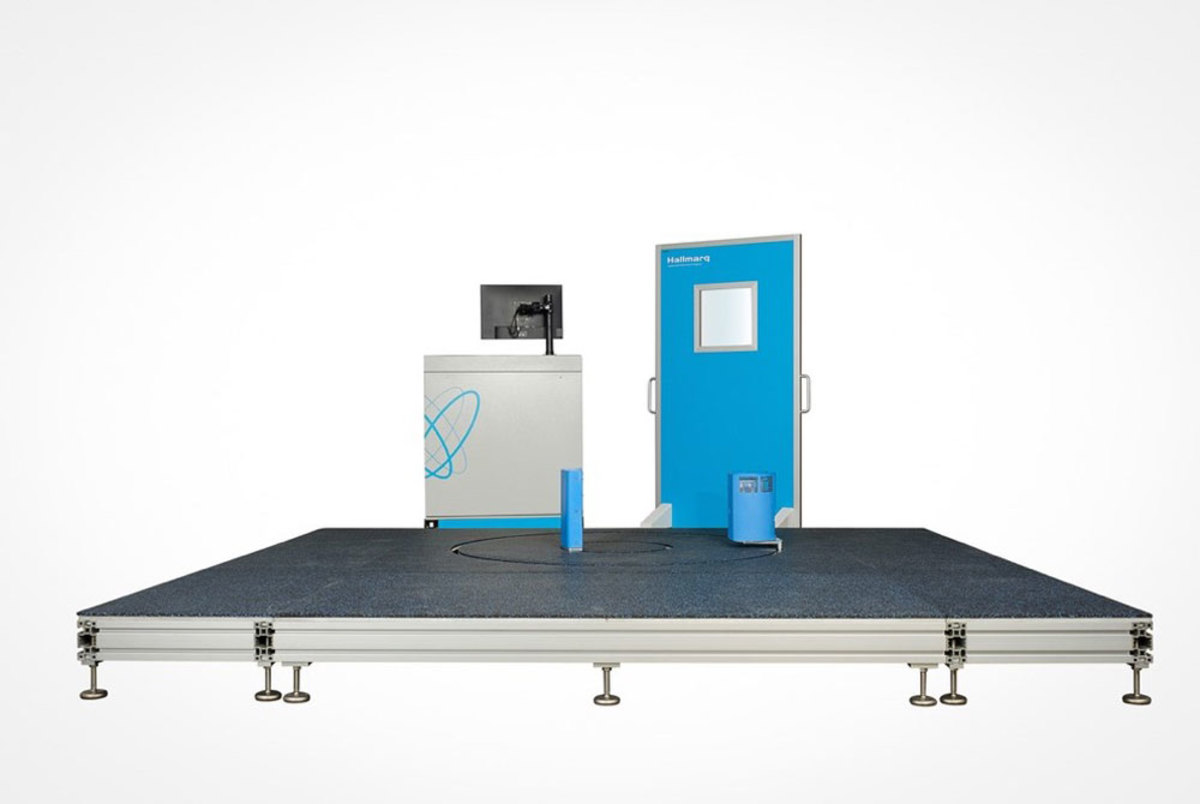
“Just like our Standing MRI, our Standing Equine Leg CT is designed from the ground up with the horse in mind, so it’s compact, open and safe for the horse and ideal for today’s equine vet who requires visualization of bone and potentially cartilage,” said Nick Bolas, founding director at Hallmarq. He explained that the system uses a novel dual concentric ring design, which enables the detector plate to remain very close to the region of interest, thereby improving image quality.
Bolas said the Standing Equine Leg CT is a good fit for veterinary practices wanting to step up to 3D imaging in the evaluation of their lameness cases. Additionally, Hallmarq is one of the few companies to incorporate motion correction technology to better ensure high-quality, clear images in the standing patient.
“We’ve learned over the years of working closely with horses and their vets, that when you can keep a horse standing under light sedation, it’s better for the horse and is much more acceptable to the owner,” said Jos Belgrave, Hallmarq’s CEO.
Hallmarq’s dedicated in-house team, including Bolas, has been working in collaboration with several equine clinics to image live horses and to ensure that the system meets the image quality, robustness and efficiency levels required for use in clinical practice.
As with their other systems, Hallmarq considers the practices’ total cost of ownership in its design decisions, providing a solution that is accessible to most clinics. Cone beam technology (CBCT) is ideal for distal limb imaging since it can detect non-displaced fractures, subtle changes in bone density and small osteophytic lesions without the expense, radiation risks and 3-phase power requirements of fan beam systems.
Having successfully scanned over 65 live, standing patients with the new CT, Belgrave said “several features and benefits have proven indispensable,” including:
- Fast, 3D imaging with 60-second scan times
- Exclusive motion correction technology
- Simple user interface and easy-to-use system
- Small footprint with an open design for patient safety
- No anesthesia costs or additional staffing needed
- Profitability with just 10-15 cases per month, depending on clinic and geography
- Fully backed by Hallmarq’s Q-care program, providing training, maintenance and system support
Hallmarq will include the slCT in their virtual booth during the AAEP meeting so veterinarians can find out about this new system and discover how it might fit with their practice.
Hallmarq Veterinary Imaging was founded 20 years ago by horse-owner Nick Bolas, who wanted to develop a diagnostic imaging system that would assist veterinarians to accurately diagnose a horse’s injury. He, and a team of imaging experts, many of whom remain with the company to this day, worked with local vets in the South-East of the UK, to develop one of the first MRI units for horses. Since then, Hallmarq has continued to improve MRI technology for horses and has gone on to develop a veterinary-specific, small animal MRI system, using expertise from across the company.
About Hallmarq Veterinary Imaging
Hallmarq Veterinary Imaging is an award-winning global leader in innovative animal-specific diagnostic imaging solutions. As industry specialists, we focus on providing safe, convenient and affordable advanced imaging for practicing veterinarians worldwide. With a shared passion for improving animal health, we’ve partnered with our customers since 2000 to support over 240,000 equine scans in 25 countries. Our exclusive dedication to the industry and exceptional support we provide our customers has resulted in increased access to advanced clinical systems for small animal and equine practices around the globe. Hallmarq Veterinary Imaging Ltd. has offices in the U.K. and North America. Find out more at www.hallmarq.net