Corneal ulcers are common in horses, and uncomplicated cases should heal within one to two weeks. Nonhealing or recurrent ulcers require a closer look at the underlying cause, which can include eyelid abnormalities, foreign bodies, tear film abnormalities, infection, edema, eosinophilic keratitis, corneal mineralization, corneal dystrophy, and more.
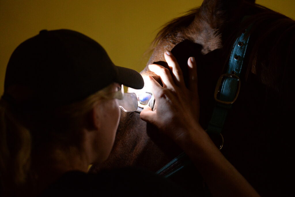
During a presentation at the 2025 British Equine Veterinary Association Congress, Samantha Dixon, BVM&S, Cert(AVP), MANZCVS(EqMed), MRCVS, of Optivet Equine, in Portsmouth, U.K., explained how to diagnose some of the more unusual cases.
Examining Corneal Ulcers
A thorough clinical examination can provide veterinarians with a wealth of information about a corneal ulcer. Dixon recommended taking a systematic approach to the ocular exam, starting with the eyelids. Can the horse blink? Does he have neurologic issues? Are there masses, foreign bodies, aberrant hairs, or cilia causing trauma?
Then move on to the tear film. Dixon suggested using a Schirmer tear test to determine tear quantity, with > 15-20 millimeters/minute being normal. Also look at tear quality: Is the cornea shiny and moist or matte?
Assess the horse’s pain, she said, and whether it’s disproportionate to the size of the ulcer. Does the pupil dilate and remain dilated in response to atropine?
Lastly, look at the ulcer itself, asking the following questions:
- Where is it within the cornea?
- What shape is it?
- Are they linear?
- Are the ulcers always in the same place?
- Is there opacity in the ulcer bed?
- Is there cellular infiltrate, fibrosis, or edema?
- Are there any blood vessels, and what pattern are they in?
- Are the epithelial edges underrun, or does fluorescein bleed under the edges?
- Is there corneal edema in a wider area of the cornea?
“Then consider further diagnostic tests, of which cytology and culture are extremely important,” said Dixon. “But I would urge you to consider referring some of these cases, because it can be quite difficult to get to the bottom of the underlying cause.”
Diagnostic Tools
Fluorescein dye can help highlight areas of ulceration. It stains the stroma but not the epithelium, the Descemet’s, or the endothelium. “If the fluorescein is bleeding under the epithelial edges, that can indicate that they’re not adhered,” said Dixon. “And there are a few things that can suggest, including indolent ulcer, fungal infection, and eosinophilic keratitis.”
Most ophthalmoscopes will have a cobalt blue filter on them that can help highlight the ulceration, especially if you have small or subtle areas of uptake, she said. “Just remember that fluorescein will pool in a corneal defect, so it’s important that we’re flushing our eyes with sterile saline after staining to remove any excess fluorescein,” she added. “Then we can tell whether we have true uptake or if the fluorescein is just pooling.”
You can also use fluorescein to assess for corneal perforation with a Seidel test. “We can flood the cornea with fluorescein and get a green film on the surface,” Dixon explained. “Then if we have aqueous leakage, that will wash away the fluorescein.”
Slit lamp biomicroscopy provides you with 10-16X magnification and a bright light source to see the fine detail in the cornea. “The slit beam allows us to identify the depth of lesions within the cornea, which is particularly important with stromal loss and abscesses, but it’s also very helpful for identifying endothelial disease,” she said.
Cytology and culture are very important diagnostic tools and, in Dixon’s opinion, potentially underused. She offered tips for making the most of your cytology and culture:
- The cornea must be fluorescein positive.
- Always do cytology with culture. “Cytology will help you interpret culture results,” she said. “So if you’re suspicious of a bacterial infection and you’ve cultured a bacteria, you want to know that you’ve got neutrophils and intracellular bacteria to support your diagnosis.” Without cytology, you might miss things like fungal hyphae and eosinophilic keratitis, she added.
- If possible, try to make a cytology preparation on a slide and send it to the lab in addition to the swab for culture. Dixon said this tends to give you a better sample than leaving it to the lab to make their own cytology preparation from the swab you’ve submitted.
- Use a Cytobrush, if possible.
- Sample the epithelial edges in addition to the ulcer bed because the highest load of infectious organisms can usually be found in and around the edges.
- Charcoal for culture and sensitivity.
- Contrary to what some reports say, fluorescein and tetracaine do not prevent sampling.
Dixon then explained how to diagnose five unusual causes of corneal ulcers.
Fungal Keratitis
Superficial fungal keratitis typically affects the epithelial and subepithelial layers of the cornea. These are sometimes referred as uveitis because the horse will have a very painful eye with myosis and not much else to see.
“But if we do a very thorough clinical exam, we can see these kind of sparkly epithelial erosions, often linear and branching,” said Dixon. “These will variably uptake fluorescein.”
Diagnosis includes cytology and culture, clinical appearance, and response to antifungal treatment. The main differentials for fungal keratitis are viral or dendritic ulcers, she noted.
Fungal Stromal Abscess
“Fungi are ubiquitous in the equine environment and part of the commensal ocular flora,” Dixon explained. “So we just need disruption of our epithelial barrier, and fungi can invade the stroma.”
Hot and humid weather as well as a history of recent topical steroids or antibiotics can predispose a horse to fungal abscesses. Key clinical features include:
- Very painful.
- Can have severe secondary uveitis.
- Gray/white (can also be yellow) cellular infiltrate in the ulcer bed.
- Inhibition of angiogenesis (won’t see any blood vessels).
- Keratomalacia.
- Irregular texture to ulcer bed (an “icing sugar” appearance).
- Thickened or underrun edges.
- Deep furrows at the edges of the ulcer.
To diagnose fungal infections, use clinical appearance and history as well as cytology to identify the fungal hyphae. If you have the ability to do a cytology in-house, you can get a result the same day—or the next day if you send it to a lab.
Don’t rely on fungal culture, Dixon warned, which takes up to two weeks to get a result. “Having said that, you don’t always manage to find your fungus on cytology, so if you’re suspicious of a fungal abscess, treat for fungus even if your cytology hasn’t given you back fungal hyphae,” she said.
Burdock Keratitis/Foreign Bodies
Burdock is a plant native to Europe and Asia that has burrs that can get tangled in horses’ manes and forelocks.
“The seedheads dry out and split, and you get these tiny, microscopic, barbed bristles that make excellent ocular foreign bodies,” said Dixon. “The barbed bristles lodge in the palpebral conjunctiva and cause persistent corneal trauma.”
In the U.K., this is a seasonal diagnosis generally seen from October to December. The condition has also been described in the United States. A typical history is a nonhealing ulcer with a very painful eye or an ulcer that has healed but ocular pain persists, particularly in horses that have had burrs in their forelocks, she said. Key characteristics include:
- Located in the palpebral conjunctiva, primarily in the upper eyelid and 5-8 millimeters from the eyelid margin.
- Small, pale nodule of inflammatory tissue surrounding the foreign body.
- Ulcers are often linear.
- Focal corneal fibrosis, edema, and focal vascularization.
- Can sometimes see scratches in the ulcer bed.
For diagnosis, use history and a careful clinical examination of the eyelids with bright light and magnification—preferably a slit lamp, said Dixon. She said these foreign bodies are very difficult to see, even with a slit lamp, but coloring the eyelids in with a surgical marker or fluorescein can help. Fortunately, she added, they’re easy to remove via conjunctivectomy.
The main differential for burdock keratitis is ectopic cilia.
Endotheliitis
Endotheliitis is inflammation of the corneal endothelium characterized by keratic precipitates (inflammatory cells on the corneal endothelium). They inhibit the normal corneal endothelial pump function, causing corneal edema that leads to recurrent ulceration.
“The typical history here is recurrent ulcers that heal but edema remains,” said Dixon. “Often when you quiz the owners, the cloudy spot of edema preceded the ulcer. And usually, the area of edema is wider than the ulcer. They’re often focal in the early stages and can be ventral in the cornea. They can progress, and we can get an increase in keratic precipitates, progressive edema, and endothelial pump loss. We can see uveitis with these, and some progress to glaucoma.”
Slit lamp examination is key to diagnosing endotheliitis, because you can see the keratic precipitates on the endothelium very clearly. Tonometry is helpful to know if the horse has glaucoma or significant uveitis.
A subset of these cases, known as heterochromic idridocyclitis and keratitis (HIK), causes a much more severe form of endotheliitis often accompanied by significant uveitis, said Dixon. Clinical features of HIK include:
- Corneal edema, usually ventral and rapidly progressive.
- Darkly pigmented keratic precipitates.
- Iris depigmentation.
- Uveitis.
- Poor response to treatment.
- Second-degree glaucoma.
Eosinophilic Keratitis
Eosinophilic keratitis is a seasonal (summer) diagnosis believed to be a hypersensitivity or immune-mediated reaction to parasites or allergens. Dixon said it’s characterized by eosinophilic infiltration into the cornea and recurrent or nonhealing ulcers. Clinical features include:
- White/gray/yellow plaques on the cornea with a “ground glass” appearance to the ulcer bed.
- Nonhealing ulcers that often have underrun edges.
- Ulcers that are perilimbal, which is an otherwise unusual location.
- Multiple ulcers per eye.
- Bilateral.
- Caseous discharge.
- Variable pain and vascularization.
- Commonly, secondary infection.
“Again, this is a cytological diagnosis, so we need to be doing cytology in these cases and identifying eosinophils on our cytology,” said Dixon. “We might also see neutrophils if we’ve got second-degree infection.” The main differential is fungal keratitis.
Related Reading
- Corneal Cross-Linking to Treat Ulcerative Keratitis in Horses
- Tips for Enucleating Horses in the Field
- Prepurchase Ophthalmic Examinations: What Do You Need?
Stay in the know! Sign up for EquiManagement’s FREE weekly newsletters to get the latest equine research, disease alerts, and vet practice updates delivered straight to your inbox.