In this episode of the Disease Du Jour podcast, Lauren Schnabel, DVM, PhD, DACVS, DACVSMR, joined us to discuss cellulitis and lymphangitis in horses. She explained how she differentiates the two conditions, her diagnostic approach in the field and clinic, her preferred treatment strategies and adjunctive therapies, and more.
This episode of Disease Du Jour is brought to you by Equithrive.
How to Differentiate Cellulitis and Lymphangitis
Schnabel explained that cellulitis is inflammation or infection of the horse’s subcutaneous tissue, while lymphangitis refers to inflammation or infection of the lymphatic vessels. Lymphangitis, which usually affects the hind limbs, often presents with marked inflammation; the horse might have a “tree trunk” limb that’s inflamed from the coronary band to the top of the leg. In severe cases, the horse might leak lymphatic fluid.
Cellulitis is often associated with a wound or abrasion. While it sometimes looks similar to lymphangitis, it can be less severe and might only affect the distal limb.
“When it’s more minor and starting on the distal limb or there’s a wound involved, then we’re thinking more cellulitis,” Schnabel said. “But where things get tricky is if cellulitis progresses, they can look very similar. And cellulitis can also lead to lymphangitis. If you have a horrible cellulitis that’s left untreated for a time or not responding appropriately to treatment, then the lymphatic vessels can actually scar as part of that process, and the horse can be prone to lymphangitis in the future.”
Schnabel said the horse’s history and breed (drafts and cobs are predisposed to lymphangitis, and lymphangitis is more likely to reoccur) can also help you differentiate between the two conditions.
Diagnosing Cellulitis and Lymphangitis
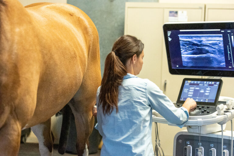
Schnabel’s diagnostic approach involves taking radiographs to rule out an insidious bone pathology or foreign body and then using ultrasound.
“The cellulitis cases are going to have very thick subcutaneous tissues with fluid in them, and they can also get focal abscessation or abscess pockets under the skin that need to be opened and drained in the more severe cellulitis cases,” she said. “The lymphangitis cases are going to have very dilated lymphatic vessels.”
In hospital settings, Schnabel sometimes uses lymphoscintigraphy to visualize the lymphatic vessels in horses with chronic lymphangitis. This technique is similar to bone scan for orthopedic cases, but it uses a different radioisotope (technetium-99m sulfur colloid).
Systemic and Adjunctive Treatment Options
For typical cellulitis cases, Schnabel’s mainstay treatment approach is regional limb perfusion, followed by firm compression bandages and icing. Uncomplicated cellulitis cases that are caught early typically do well and don’t cause further issues, she said.
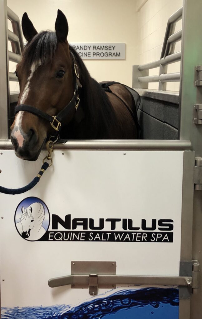
For severe cellulitis, Schnabel puts the horse on IV antibiotics and tries to reduce swelling until she can access a vein for regional limb perfusion. Her clinic has a saltwater spa, which helps reduce edema. When the horse is out of the spa, she focuses on bandaging to further reduce swelling.
Horses with lymphangitis also do well in the spa, she said. These cases can be a bit more complicated, because bandaging puts static pressure on the leg but doesn’t improve lymphatic flow. “If the horses are painful, they can’t be walking around very much in that state, but you need them moving to generate fluid up the limb,” she said.
Schnabel co-founded the company Vetletics, which designed the EQ Press, a dynamic compression device that helps improve lymphatic flow. The device goes around the horse’s legs and fills with air in sequential chambers to push fluid from the coronary band up the leg. “We did lymphoscintigraphy with or without the EQ Press on, and it dramatically accelerated lymphatic clearance,” she said, noting that the study was not performed on horses with damaged lymphatics.
Listen to the podcast episode to learn more, including the role of bacterial culture and sensitivity testing, potential secondary complications, and early prognostic indicators.
About Dr. Lauren Schnabel
Lauren Schnabel, DVM, PhD, DACVS, DACVSMR, is a Professor of Equine Orthopedic Surgery in the Department of Clinical Sciences at NC State College of Veterinary Medicine. She completed her DVM, Large Animal Surgery Residency, and PhD at Cornell University. She is board-certified in both the American College of Veterinary Surgery and the American College of Veterinary Sports Medicine and Rehabilitation. Schnabel’s research focuses on stem cell immunology, use of biologic therapies to treat musculoskeletal injuries and diseases, and advancing equine rehabilitation protocols.
Related Reading
- Disease Du Jour: Regional Limb Perfusions
- Mastering Regional Limb Perfusions in Horses
- AAEP Health Coverage: Pneumatic Compression Therapy Device for Horses
Stay in the know! Sign up for EquiManagement’s FREE weekly newsletters to get the latest equine research, disease alerts, and vet practice updates delivered straight to your inbox.
![[Aggregator] Downloaded image for imported item #19998](https://s3.amazonaws.com/wp-s3-equimanagement.com/wp-content/uploads/2026/05/29122748/EDCC-Unbranded-5-scaled-1-768x512.jpeg)