Butterfly Network is creating an Ultrasound Reference Library to assist equine practitioners better use point-of-care ultrasound (Butterfly iQ+ Vet). This page is focused on Palmar Metacarpal Soft Tissues, with information from Cooper Williams, VMD, DACVSMR, Certified ISELP instructor. (For more information on Williams see his bio below.)
Brought to you by Butterfly iQ+ Vet
Palmar Metacarpal Soft Tissues
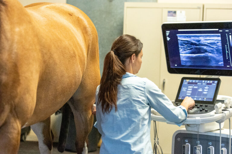
Other than the reproductive tract, the most common region evaluated in equine sonography is the palmar metacarpal soft tissue region. This is a great place to start with your Butterfly iQ+ Vet. Pull the probe out and document your soft tissue clinical findings!
Always start with good practices of preparation. If possible, clip the limb! This can’t be said with enough emphasis. The Butterfly IQ+ Vet works in many areas of the body with the use of alcohol alone and without removing the hair. However, the subtleties of the tissues especially superficially, are best seen with clipping, hot water, and the use of ultrasound gel. A standoff pad is also recommended, allowing manipulation of the area of interest into an optimal focal zone. The standoff also removes or reduces the reverberations in the area of interest, improving image quality. Ultrasound gel is a must for optimal transmission and should be placed both inside and outside on the standoff pad. This allows smooth gliding for the real time video clips as well.
It is important to perform sequential images throughout the palmar metacarpal region. The author uses the zone system (Z1A, 1B, 2A, 2B, 3A, 3B and 3C) to perform a minimum of seven frozen images distributed evenly through the metacarpal region (starting proximally). This allows for methodical standardization of imaging, especially important when sharing images with other practitioners. A transectional (cross section) and longitudinal image are performed in each zone.
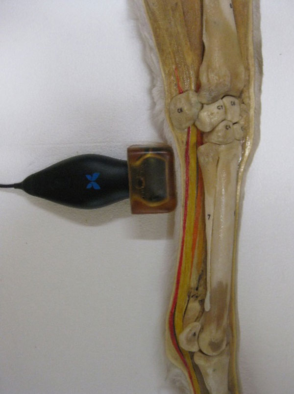
Image standardization through adherence to ultrasound convention is also very important. This refers to organization of images on the screen and international convention dictates placing medial on the left side of the screen, proximal on the left, dorsal on the left, cranial on the left and when comparing sides with a split screen, left on the left. To date, not all in the profession have accepted these recommendations. After acquisition of still images, the author recommends performing a real time running video clip proximally to distally through the same region. There are a few more advanced tools that can be used to extract more information from this region (i.e., angle contrast imaging, lateral imaging, flexed imaging, etc.) and we will elucidate those in future reviews.
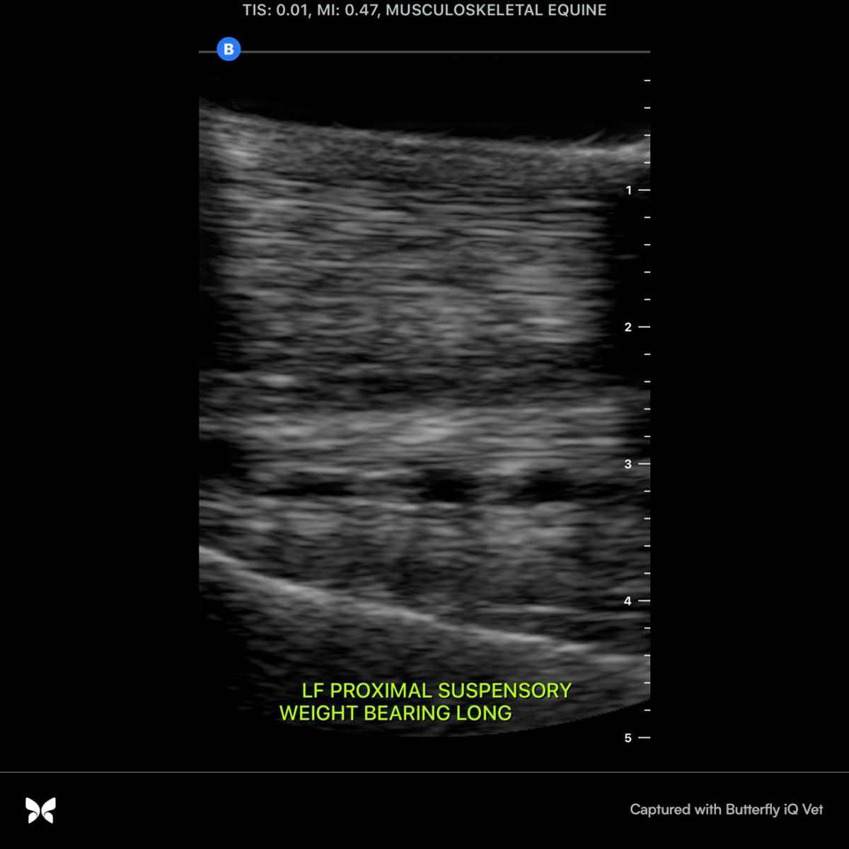
The structures portrayed in this proximal palmar metacarpal image above, from top to bottom, are the superficial digital flexor tendon, the deep digital flexor tendon, the accessory ligament to the deep (also known as the inferior check ligament) and the suspensory ligament. The depth has been adjusted to approximately 1cm beyond the bone surface, the bright white line at the base of the image is the palmar aspect of MCIII (the cannon bone) bringing the suspensory ligament into a more focused part of the image.
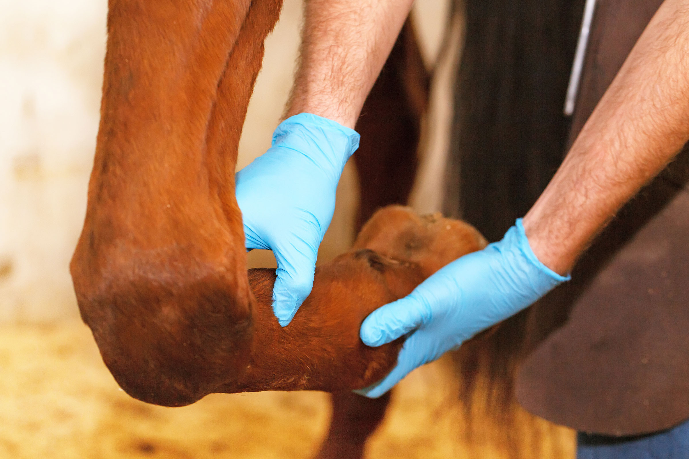
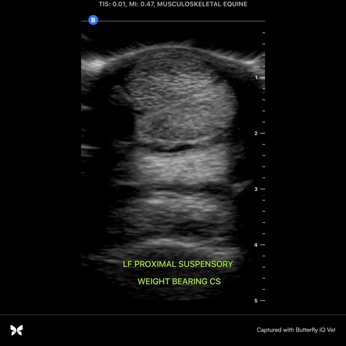
Because the narrowest part of the palmar metacarpal soft tissues is the most palmar aspect, air artifact is created through the deeper and wider accessory ligament of the deep digital flexor tendon and suspensory ligament. Due to this artifact created in the standing limb, accurate measurements are not possible. Two tools allow complete imaging of these structures. First, the limb is held flexed and scanned in this position. When flexed, the flexor tendons move to a medial/lateral position, forming a natural standoff for the deeper structures. Second, the limb is scanned from the lateral aspect of the standing limb, directly imaging the accessory ligament of the deep digital flexor tendon in full cross section. Again, these tools will be discussed in depth, in future reviews. This cross section is in zone 1B and will be followed below with an image acquired in longitudinal orientation in the same zone.
This zone is the metacarpal portion of the proximal enthesis of the suspensory ligament. It is equally important to evaluate soft tissue structures and their entheses (bone origins and insertions). Ultrasound is the most sensitive tool we have in the field for bone surface detail. The Butterfly iQ+ Vet makes it easy to have a readily available and easy-to-use tool for this. This image shows a healthy smooth white line of bone surface and normal ligamentous fibers emerging from the enthesis. An abnormal image might elucidate small enthesophytes or even avulsions of bone. A more thorough discussion regarding the suspensory ligament and its enthesis in both forelimbs and hindlimbs will also be the subject of a future review.
Brought to you by Butterfly iQ+ Vet
Cooper Williams, VMD, DACVSMR, Certified ISELP Instructor
Dr. Cooper Williams grew up in Cecil County Maryland, where he learned to play polo and train polo ponies with his family. He ultimately obtained a five goal rating and played professionally for five years. In the summer of 1981, he was a member of the United States polo team that competed throughout England. He coached the Valley Forge Military Academy polo team from 1981 to 1985. In 1984, he graduated with honors from the University of Pennsylvania School of Veterinary Medicine. He accepted a post graduate internship at Delaware Equine Center, where he developed their ultrasound imaging program. In 1987, he moved to Maryland where he worked for two years at Maryland Equine Center, also developing their ultrasound program. In 1989, Williams started his own practice, focusing on diagnostics and sports medicine in equine athletes. After three years of rigorous study, learning in depth about advanced diagnostic imaging, Williams passed his examination and was certified by the International Society of Equine Locomotor Pathology (ISELP) in 2009 and has continued on as a certified instructor with ISELP, teaching other equine veterinarians all over the world. Cooper is one of a small group of veterinarians in the world who is certified by the International Society of Equine Locomotor Pathology in advanced ultrasound imaging. In August 2014, Williams successfully passed his examination and is officially a Diplomate of the American College of Veterinary Sports Medicine and Rehabilitation. There are only a small group of veterinarians in the world that now can claim they are a specialist in sports medicine and rehabilitation. He is an active member of the North American Regenerative Medicine Association as well as being on the veterinary advisory board for ACell, Inc., a tissue engineering company.
Williams is a paid advisor for Butterfly Network.
![[Aggregator] Downloaded image for imported item #19998](https://s3.amazonaws.com/wp-s3-equimanagement.com/wp-content/uploads/2026/05/29122748/EDCC-Unbranded-5-scaled-1-768x512.jpeg)