Fractures are a common occurrence in horses of all ages and breeds, but the development of various imaging modalities over the past 10 years is helping significantly to advance management techniques. The Equine Veterinary Journal’s (EVJ) new online collection, with an introduction from imaging expert Renate Weller, new President of the British Equine Veterinary Association, showcases the diagnostic imaging breakthroughs that are helping to identify causes, support prevention and improve fracture outcomes.
Fractures are often associated with complications and a poor outcome. In “Epidemiology of fractures: the role of kick injuries in equine fractures,” the authors conclude that kicks are the most common cause, most often seen in ponies and with mares being at higher risk.
Medial intercondylar eminence of the tibia (MIECT) fractures are also usually associated with a traumatic event, concluded the study “Fractures of the media intercondylar eminence of the tibia in horses treated by arthroscopic fragment removal.” Rarely reported in horses, they need prompt diagnosis and arthroscopic removal of the fracture for the best prognosis.
Stress fractures are the leading cause of lost training days and a common cause of euthanasia in racehorses. “Analysis of stress fractures associated with lameness in Thoroughbred flat racehorses training on different track surfaces undergoing nuclear scintigraphic examination” showed a higher incidence of stress fractures in horses training on synthetic surfaces, but other factors such as training philosophy are also likely to be of influence.
“A longitudinal study of fractures in 1488 Thoroughbred racehorses receiving intrasynovial medication” has addressed the shortfall of knowledge on the association between intrasynovial medication and fracture risk. The study concluded that 3% of horses suffered serious injury following medication and that greater use of pre-medication diagnostic imaging may reduce injury rates.
Condylar fractures of the distal metacarpus are an important cause of fatality in Thoroughbred racehorses. “Unicortical condylar fracture of the Thoroughbred fetlock” concludes that veterinary vigilance and timely intervention may considerably reduce the risk of catastrophic injury during racing.
Increased subchrondral bone plate thickness at the parasagittal groove can be a useful indicator of lateral condylar fracture concludes the study “Can we use subchondral bone thickness on high-field magnetic resonance images to identify Thoroughbred racehorses at risk of catastrophic lateral condylar fracture.”
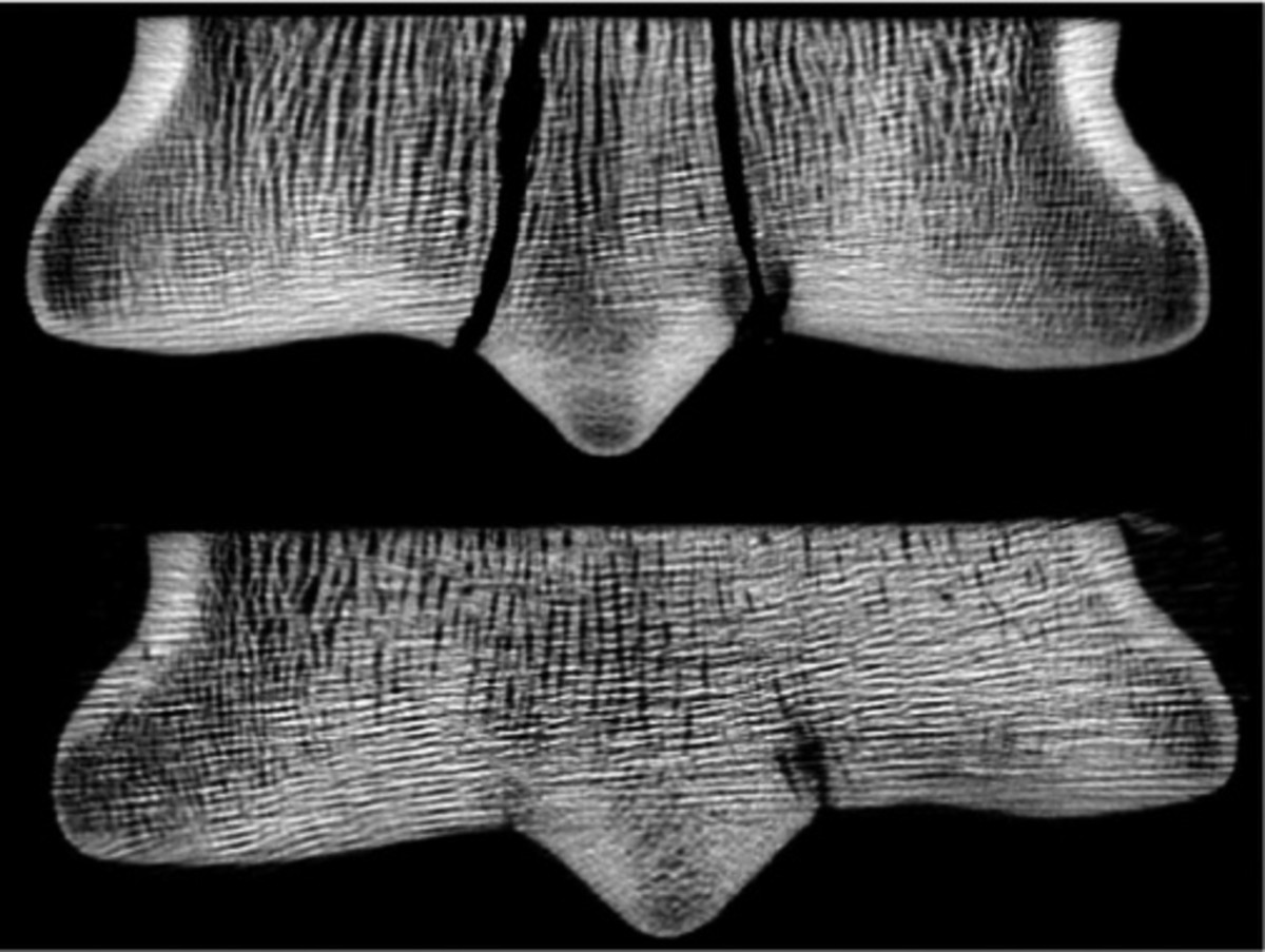
Micro CT can also demonstrate fatigue damage in the distal metacarpal subchondral bone. The study “Subchrondral bone microdamage accumulation in distal metacarpus of Thoroughbred racehorses” concludes that reduced intensity of training and increased rest periods may limit microdamage accumulation.
However, high resolution peripheral quantitative CT does not specifically predict condylar fracture concludes the study “Can high resolution peripheral quantitative computed tomography imaging of subchrondral and cortical bone predict condylar fracture in Thoroughbred racehorses?” This may be because there is a lack of clear distinction between the subchrondral plate and the trabecular bone and because measurement of the subchrondral bone thickness is complicated by adjacent palmar osteochrondral disease lesions.
In terms of fracture configuration, diagnosis method of repair and outcome the study “Short frontal plane fractures involving the dorsoproximal articular surface of the proximal phalanx: description of the injury and a technique for repair” showed that these were more common in hind limbs. Fractures exiting the bone distal to the metacarpophalangeal/metatarsophalangeal joint capsule can often be minimally invasively repaired and carry a good prognosis for a return to work.
“Central tarsal bone fractures in horses not used for racing: computed tomographic configuration and long-term outcome of lag screw fixation” showed that fractures in non racehorses had a distinct configuration but that subtle additional fractures lines can occur, likely a result of chronic stress. Accurate diagnosis of the fracture configuration resulted in successful internal fixation and a very good prognosis.
A successful technique for the repair of slab fractures was shown in the study “Slab fractures of the third tarsal bone: minimally invasive repair using a single 3.5 mm cortex screw placed in lag fashion in 17 Thoroughbred racehorses.” The study concluded that surgical repair is a viable alternative to conservative management.
Post-operative imaging is mandatory for the assessment of repair and any potential complications and in human medicine MRI is the preferred modality. “Magnetic resonance imaging of an equine fracture model containing stainless steel metal implants” showed that the multiacquistion variable resonance image combination (MAVRIC) significantly reduced the artifacts created by metallic implants and improved anatomic delineation.
“The advancement of imaging modalities over the past ten years has significantly improved the management of equine fractures,” said Celia Marr, editor of the EVJ. The articles included in this fascinating collection not only highlight the current scope of diagnostic imaging for a range of equine musculoskletal conditions but also clearly indicate its potential in continuing to improve clinical assessments, treatment and outcomes.”
The new collection is available free online at https://bit.ly/2zQHKCp.