In this episode of the Disease Du Jour podcast, Elsa Ludwig, DVM, PhD, MS, CVA, DACVS (LA), discussed melanoma in horses and a novel technology she has been using to treat skin tumors called H-FIRE.
This episode of Disease Du Jour is brought to you by Bimeda.
Prevalence and Location of Melanoma Tumors
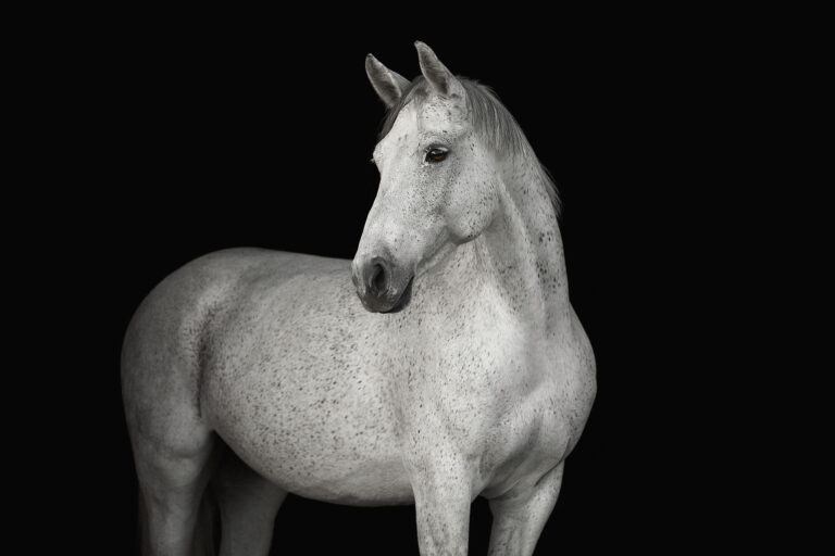
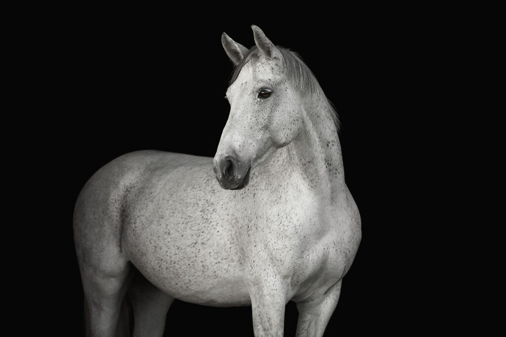
Ludwig explained that melanomas comprise 6-15% of all equine skin tumors and affect up to 80% of older gray horses.
Tumors can develop anywhere on the horse, but the most common locations are the underside of the tail, the perineum, and the external genitalia. Other common locations include the parotid region, commissures of the lips, around the eyes, and within the crest of the neck, she explained. Tumors can also hide in the guttural pouches, within the sheath, or alongside the rectum.
Evaluating Melanomas
“[Melanomas] are fairly easy to diagnose. Because of the gray horse color, we can typically assume it’s a melanoma,” Ludwig said. “However, when I get a horse at my hospital for the first time, I do a really thorough exam because I am looking for hidden melanomas.”
Ludwig’s evaluation includes palpation of all visible surfaces as well as a sedated rectal palpation. She employs rectal ultrasound to evaluate for perirectal tumors and deeper tumors within the pelvic region. She also performs a thoracic and abdominal ultrasound, looking at the horse’s lungs and abdominal organs.
Histopathology of tumor biopsies confirms the diagnosis and differentiates between different melanoma types, which include:
- Naevus: The only truly benign melanoma type, which has limited epidermal or superficial dermal involvement.
- Dermal melanoma: Isolated, discrete, spherical masses typical of young to older gray horses. They are often benign but can become malignant.
- Dermal melanomatosis: Multiple coalescing masses, typically affecting older horses. They are also often benign but can become malignant.
- Anaplastic malignant melanoma: The most aggressive form of melanoma, which grows rapidly and frequently metastasizes to other organs. This type of melanoma is rare and typically affects geriatric and non-gray horses.
“Ninety-five percent of tumors are considered benign, especially when we catch them early on,” Ludwig said. “However, up to 30%, and sometimes in some reports even higher, can advance into the type of melanoma that becomes malignant, and then they ultimately metastasize.”
Leave Alone or Treat?
Because most types of melanomas have the potential to become malignant and metastasize, Ludwig typically recommends removing tumors.
“It’s really hard for us to tell just by looking at them,” she said. “You can’t just grossly say, ‘That little one’s going to be fine, and that little one’s not.'”
However, she does not always recommend treatment for horses with advanced anaplastic tumors affecting significant structures, because these horses usually die within a few months.
Treatment options for melanoma include surgical resection, CO2 laser ablation, cryosurgery, or a novel technique called high-frequency irreversible electroporation (H-FIRE). Other options that have been used historically with unreliable results include cimetidine, Oncept (the canine melanoma vaccine), and autologous vaccines using samples of the horse’s tumor.
Chemotherapy is another traditional treatment, but it poses risks to the veterinarian handling the drug and anyone who touches the horse after treatment. “I don’t use it very often for treatment anymore in melanomas because I have access to other treatment options,” Ludwig said.
H-FIRE
Ludwig uses a relatively novel treatment modality for treating skin tumors, including melanoma, called H-FIRE. It was originally developed at Virginia Tech by Dr. Mike Sano, who brought the technology with him when he moved to North Carolina State University.
“The general technology of electroporation has been around for a while,” she said. “The concept behind it is the application of electrical pulses to those tumor tissues. And what this does is increase the membrane permeability of those locally exposed cells, and it also helps induce cell death.”
Historically, available machines required horses to be placed under general anesthesia because the technique caused significant muscle contractions. H-FIRE has helped mitigate this limitation.
“The technology uses pulsed electrical fields rather than a continuous electrical field, and this helps increase the transmembrane potential of those tumor cells,” she explained. “So it pokes holes essentially in the cell walls, and that causes the cells to rupture and die.”
This technology eliminates the intensity of the muscle contractions that can occur with electroporation, Ludwig said, negating the need for general anesthesia. H-FIRE also induces a local immune system response, which improves antigen presentation and adaptive immune response and promotes anti-tumor activity.
Ludwig said the number of treatments required varies. Moderately sized or smaller nodules usually resolve after one treatment, while large or coalescing tumors can require one to four treatments. She employs a combined treatment approach on most patients, using surgical or laser excision for some masses and H-FIRE for others.
Listen to the podcast episode to learn more about H-FIRE.
About Dr. Elsa Ludwig
Elsa Ludwig, DVM, PhD, MS, CVA, DACVS (LA), is a clinical assistant professor of equine surgery at Virginia Tech’s Marion duPont Scott Equine Medical Center. She is an equine soft tissue surgeon who specializes in colic, tumor removal, oral and maxillofacial surgery, sinus infection treatment, and complex dental surgery.
Ludwig holds a DVM degree from the University of Wisconsin-Madison School of Veterinary Medicine, a Master of Science focusing on equine septic arthritis from the Virginia–Maryland College of Veterinary Medicine, and a PhD in comparative biomedical science from North Carolina State University.
Board-certified by the American College of Veterinary Surgeons-Large Animal and a Certified Veterinary Acupuncturist, Ludwig has served as an equine soft tissue surgery clinician, emergency clinician, and locum surgeon at institutions including the veterinary college at Virginia Tech and N.C. State University.
Related Reading
- Golden Rules for Removing Equine Skin Tumors
- Treatment Outcomes for Perianal Melanoma Removal in Horses
- Disease Du Jour: Ocular Squamous Cell Carcinoma
Stay in the know! Sign up for EquiManagement’s FREE weekly newsletters to get the latest equine research, disease alerts, and vet practice updates delivered straight to your inbox.