It is unfortunate that everyone reading this article couldn’t have attended this Focus meeting. While we have provided—with the support of Soft-Ride—some key points from this conference, there was much more of interest to equine veterinarians. Check out the feature article from the meeting titled” AAEP 2019 Summer Focus Conference.”
Following are a few tidbits and take-homes from other talks.
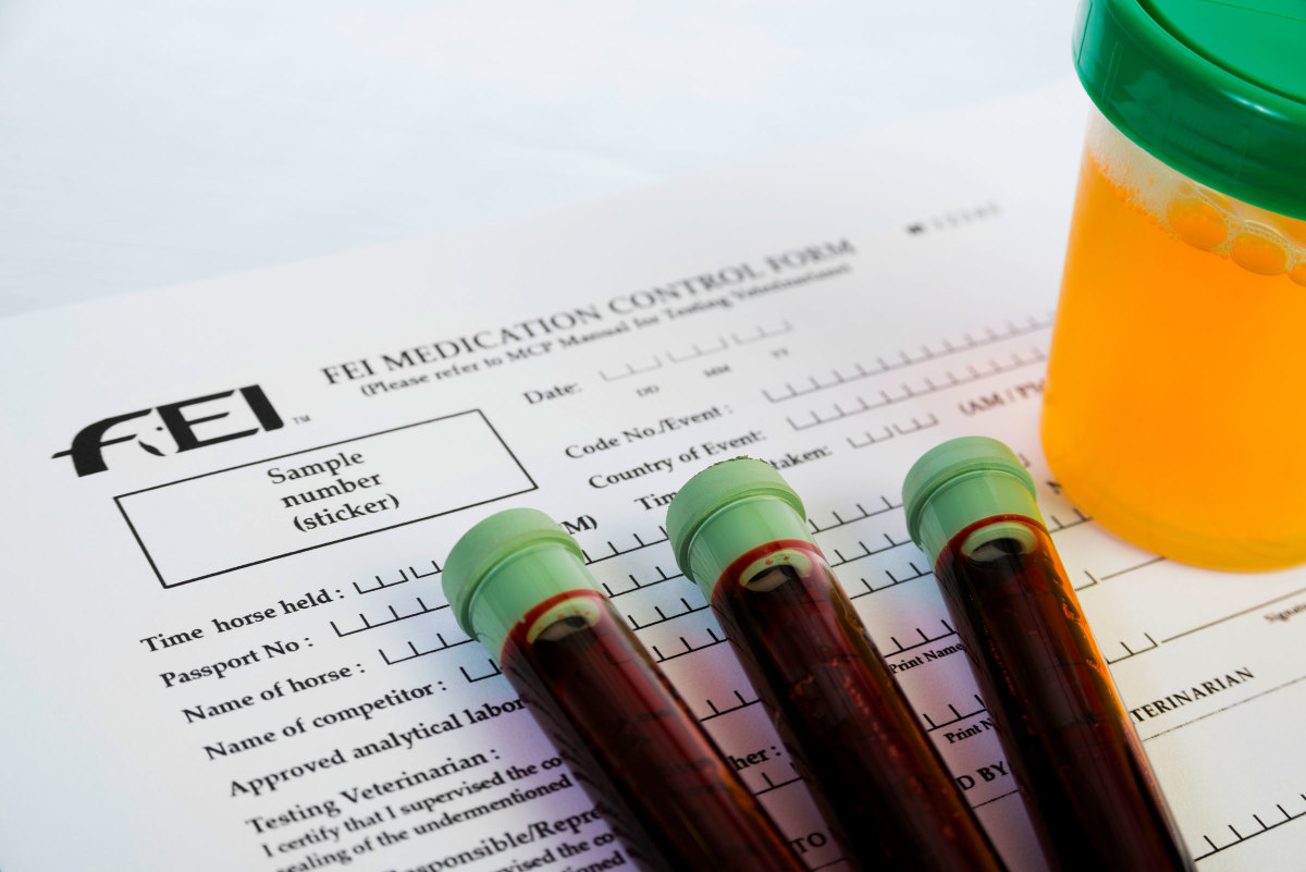
Katie Seabaugh, DVM, DACVS, DACVSMR, of CSU, spoke on “Managing Horses under USEF and FEI Rules.” Her takeaway was: “A final recommendation regarding the management of performance horses is to know the current rules and DOCUMENT, DOCUMENT, DOCUMENT. In the unfortunate case that a horse tests positive for a banned or prohibited substance, it will be critical to have complete medical records.”
Erin K. Contino, MS, DVM, DACVSMR, of CSU, spoke on “Going the Extra Mile: Working up Cases of Poor Performance.” Her summary was that “while many overt lamenesses can be accurately diagnosed with a thorough musculoskeletal and lameness examination, diagnostic analgesia and diagnostic imaging, in cases of behavioral issues and/or poor performance, traditional diagnostics may be insufficient.
“In such cases, it is encouraged to evaluate the horse under saddle, to attempt diagnostic analgesia even in the absence of overt lameness, and to evaluate the horse, rider and tack under as many circumstances as possible. Additional diagnostic tools that can be considered, as guided by the clinical examination, include imaging of the skull and axial skeleton, nuclear scintigraphy, dynamic endoscopy, gastroscopy and trial treatment with systemic medications. These cases can be time consuming and financially draining—yet equally rewarding, if a diagnosis is reached.”
Kurt Selberg, MS, DVM, MS, DACVR, of CSU, discussed “Imaging of the Foot.” In summary of his talk he said, “Using a combination of imaging diagnostics and examination approaches can help identify clinically relevant injuries to the foot. Identifying these lesions help formulate a treatment strategy and formulate a prognosis for the horses intended purpose. However, when using first-line diagnostic imaging such as radiography and ultrasound, it is important to understand the limitations and interpretation pitfalls in diagnosing injuries in the digit.”
Selberg also presented on “Imaging of the Neck and Back.” In conclusion, he said that “multiple types of pathologic changes affect the axial skeleton. Using well-positioned radiographs and detailed ultrasound exams can help localize areas of injury and help guide therapy.”
Myra F. Barrett DVM, MS, DACVR, of the CSU Department of Environmental and Radiological Health Sciences, spoke on “Multimodality Imaging of the Equine Distal Limb: Beyond the Foot.” The summary of her talk was that “in order for imaging to be a useful diagnostic modality, studies must be acquired and evaluated as a piece of the clinical picture, taking into account patient presentation, physical and lameness exam and blocking pattern. Often, more than one modality or approach is required for a complete analysis. The range of area imaged should be generous in order to avoid missing significant lesions.”
Barrett also spoke on “Diagnostic Imaging of the Equine Stifle.” Her conclusion was that “diagnosis of stiffe injury may represent a significant challenge to the equine clinician. However, a stepwise approach to the equine stifle aids in the visualization of areas where pathologic change commonly occurs and may be seen with ultrasound and radiographs. Appropriate knowledge of anatomy and common areas of pathologic change are needed to get the most out of your diagnostic imaging.”
In addition, Barrett presented on “Imaging of the Tarsus and Proximal Metatarsus.” Her conclusion was that “limitations of diagnostic analgesia for lesion localization in the tarsus and proximal metatarsus require a comprehensive approach to evaluating this area. Integrating a thorough physical examination and lameness evaluation, diagnostic analgesia and imaging is important. Often multiple imaging modalities must be utilized for the most complete diagnostic assessment.”
Colton McInturff, DVM, presented a paper titled “Equine Joint Therapies: Traditional, Biologic, and Systemic” that he and David D. Frisbie DVM, PhD, DACVS, DACVSMR, created from research done at CSU. Some of the tidbits from this presentation were:
- Autologous bone-marrow derived, culture-expanded MSCs are used most commonly in IA equine orthopedic research;
- The level of evidence supporting use of biologic therapies in the horse is currently greater for that of autologous conditioned serum (ACS) versus platelet- rich plasma (PRP);
- A 2011 survey of equine practitioners revealed that the majority of individuals (84.1%) using PSGAG administer it intramuscularly (IM). However, when comparing IA versus IM administration, it was concluded that greater potency was achieved via the IA route;
- HA has been shown to moderately decrease OA associated pain in humans and is not refuted by work conducted in equine models. There are reports of beneficial effects from intra-articular administration of combined HA and TA, and a guideline for use based on the literature is 20 to 22 mg of a mid-molecular weight HA with 3 to 5 mg of triamcinolone acetonide in a 10 to 15 mL joint as a single injection. Evidence for beneficial effects of IA HA alone does exist (2 serial injections 1 week apart), and intravenous administration of HA for prophylaxis may be advantageous.
Melinda R. Story, DVM, DACVS, DACVSMR, cVMA, cIVCA, presented on “Treating the Axial Skeleton.” Her conclusion was that “the axial skeleton is a complex and important consideration when evaluating a sporthorse for any decline in performance and changed behavior. The history, myofascial exam, lameness and neurologic exams are all important to include in the work-up. There are multiple approaches to treating these cases, and many times a multimodal approach is the most effective.”
Jackman presented a “Review of Suspensory Ligament Injury Treatment.” In his conclusion, he noted that “Suspensory desmitis is a common injury and historically the re-injury rates have been disappointing, especially in the hind limb.
A controlled rehabilitation program with serial clinical and ultrasound examinations is necessary, but additional therapies are usually needed to promote better healing.
“Extracorporeal shockwave and local injection of regenerative therapies have improved healing and increased longterm prognosis. Several surgical procedures have been described and may be indicated in more severe cases. Currently, there is not a good experimental model for equine suspensory desmitis, making direct comparisons of medical and surgical techniques difficult.”
This article and AAEP 2019 Summer Focus Conference are brought to you by Soft-Ride.