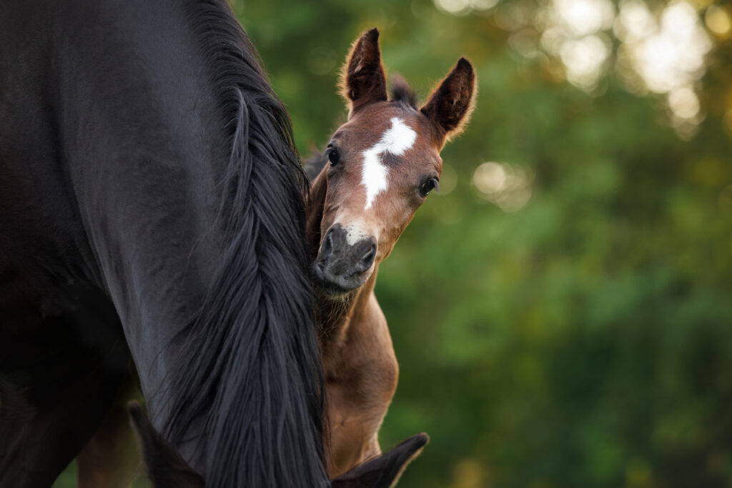
In this episode of Daily Vet Life, Kelly Knickelbein, VMD, DACVO, Assistant Professor, Section of Ophthalmology, at Cornell University College of Veterinary Medicine, introduced us to a 4-week-old Morgan colt that had been diagnosed with an advanced congenital cataract shortly after birth. He was otherwise healthy and well-bred to be a show horse.
Cataract surgery is the only vision-restoring treatment for these cases. The biggest decision, however, is whether to perform the surgery before or after weaning. Knickelbein explained that determining the timing of intervention requires balancing the potential benefits of early visual development against the increased risks associated with operating on a very young foal.
Ultimately, she and her team decided to pursue surgery pre-weaning based on the advantages of restoring vision early. She described the unique postoperative risks younger foals face, including increased susceptibility to trauma, infection, and complications related to nursing and recumbency.
Before surgery, Knickelbein and her team conducted a thorough ophthalmic and systemic evaluation. Because the dense cataract prevented visualization of the posterior segment, advanced diagnostics—including ocular ultrasonography and electroretinography (ERG)—were used to confirm retinal attachment and function. These findings, combined with an otherwise normal physical examination and bloodwork, supported the colt’s candidacy for surgery.
Knickelbein then described the phacoemulsification procedure and the delicate surgical steps required to remove the cataract and place an intraocular lens (IOL). She discussed the technical challenges of creating a successful capsulorhexis, the rationale for lens implantation, and the tradeoffs associated with IOL use in equine patients.
Although the surgery and anesthetic recovery proceeded smoothly, the most valuable lessons from the case emerged during the postoperative period.
Within days of surgery, the colt developed several complications, including a peri-incisional corneal ulcer and mild fibrin formation around the implanted lens. More significantly, he developed advanced and progressive corneal edema consistent with a suspected Descemet’s membrane detachment near the surgical incision. The condition ultimately progressed to severe bullous keratopathy, causing such extensive corneal swelling that the foal could no longer blink normally.
Knickelbein detailed the clinical decision-making that followed, including the use of a temporary tarsorrhaphy to protect the cornea and try to resolve the edema. She also noted that factors unique to young foals—such as prolonged periods spent lying on the operated side—might have contributed to the severity of the complication.
Knickelbein emphasized that successful cataract surgery extends far beyond the operating room. Careful monitoring, early recognition of complications, and prompt intervention are often the determining factors in achieving a positive outcome.
More than a year after surgery, the colt has retained his vision and continues to do well, providing a rewarding conclusion to an otherwise challenging case.
Listen to the full episode to learn how Knickelbein worked through the colt’s case from start to finish.
About Dr. Kelly Knickelbein
Kelly Knickelbein, VMD, DACVO, is a 2015 graduate of the University of Pennsylvania School of Veterinary Medicine. She completed an internship in equine medicine and surgery at Rhinebeck Equine and a comparative ophthalmology internship at the University of California-Davis. Following this, she stayed at UC Davis to complete a residency in comparative ophthalmology. Knickelbein is a diplomate of the American College of Veterinary Ophthalmologists and has been working at Cornell University since 2021. Her clinical interests include all aspects of equine ophthalmology, and her research interests include heritable ocular disorders in horses and the application of advanced ophthalmic imaging techniques in large animals.
Related Reading
- Daily Vet Life: Surgical Correction of Wry Nose
- Daily Vet Life: A Foal with Pyloric Outflow Obstruction
- Diagnosing Unusual Causes of Corneal Ulcers in Horses
Stay in the know! Sign up for EquiManagement’s FREE weekly newsletters to get the latest equine research, disease alerts, and vet practice updates delivered straight to your inbox.
![[Aggregator] Downloaded image for imported item #19769](https://s3.amazonaws.com/wp-s3-equimanagement.com/wp-content/uploads/2026/05/31141823/EDCC-Unbranded-11-scaled-1-768x512.jpeg)

