A 12-year-old Thoroughbred gelding presented with what initially appeared to be a superficial pasture injury-a wound to the lower cannon bone region near the back of the fetlock. While these types of lacerations are common, the horse’s degree of lameness raised concern for something more serious. The referring veterinarian recognized the risk associated with this anatomical location, where there is minimal tissue between the skin and critical structures like the digital flexor tendon sheath, and referred the case to Cornell University for further evaluation.
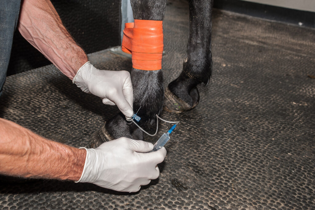
At the hospital, Rebecca McOnie, DVM, DACVS, and her team performed a thorough diagnostic workup. Initial palpation and probing of the wound were inconclusive, as was diagnostic imaging with radiographs and ultrasound due to the amount of subcutaneous gas tracking. To gain clarity, they collected synovial fluid (which appeared orange and turbid) and pressurized the tendon sheath with saline, observing fluid coming directly out of the wound.
“Based on the lack of distension of the tendon sheath and the fluid streaming out of the tendon sheath, we were able to say, alright, there’s an obvious communication here,” McOnie explained. “We got our synovial fluid analysis back as well, and those results were consistent with a septic tenosynovitis, or infection of the tendon sheath.”
With a diagnosis confirmed, the treatment plan shifted from routine wound management to aggressive intervention. The horse received local and systemic antimicrobial therapy and underwent tenoscopic lavage under general anesthesia, allowing the clinicians to flush the tendon sheath with sterile fluids while visualizing the internal structures. This approach enabled them to remove debris, look for fibrin buildup, and assess tendon integrity. Fortunately, said McOnie, they didn’t observe any significant tendon damage. They debrided the wound and performed a partial closure to allow for continued drainage.
Postoperatively, the horse received regional limb perfusions and intrathecal antibiotics and tPA (tissue plasminogen activator) to combat infection and decrease the likelihood of scar tissue or adhesion formation in the tendon sheath. Within days, said McOnie, he showed marked improvement in comfort and clinical parameters. Follow-up synovial fluid analysis confirmed that the infection was resolving, and the horse transitioned to oral antibiotics and NSAIDs before discharge. Recovery at home involved careful wound management and bandage changes and a gradual return to exercise.
This case underscores the importance of early, thorough evaluation of wounds in high-risk areas, said McOnie. Even small or seemingly superficial injuries can involve critical structures, dramatically altering prognosis and treatment requirements. Prompt recognition and appropriate referral can make the difference between a straightforward recovery and long-term complications, she said.
For a deeper dive into this case, be sure to listen to the full podcast episode.
About Dr. Rebecca McOnie
Rebecca McOnie, DVM, DACVS, earned her Doctor of Veterinary Medicine degree from the Western College of Veterinary Medicine in Saskatoon, Saskatchewan. After spending four years on the Canadian Prairies, she completed a rotating veterinary internship at Arizona Equine outside of Phoenix, Arizona. She then moved to upstate New York, where she completed a three-year residency in Large Animal Surgery at Cornell University and subsequently stayed in the role of Clinical Instructor. Dr. McOnie completed ACVS fellowship training in soft tissue minimally invasive surgery and is an Assistant Professor in the Section of Large Animal Surgery at Cornell University. Her clinical interests include orthopedic and soft tissue surgery, as well as lameness evaluation in horses and other farm animals. She is passionate about veterinary student and surgical skill education.
Related Reading
- Administration Method Does Not Affect Antibiotic Levels After IVRLP
- Disease Du Jour: Regional Limb Perfusions
- Long-Term Survival Following Synovial Lavage in Horses
Stay in the know! Sign up for EquiManagement’s FREE weekly newsletters to get the latest equine research, disease alerts, and vet practice updates delivered straight to your inbox.


![[Aggregator] Downloaded image for imported item #19998](https://s3.amazonaws.com/wp-s3-equimanagement.com/wp-content/uploads/2026/05/29122748/EDCC-Unbranded-5-scaled-1-768x512.jpeg)