To successfully obtain diagnostic images of equine mandibular cheek teeth, Amelie McAndrews, DVM, DAVDC-EQ, owner of Garden State Equine Dentistry in Princeton, New Jersey, and clinical associate at the University of Pennsylvania’s New Bolton Center, recommended preparing by:
- Using appropriate sedation (8 to 10 milligrams detomidine IV).
- Stabilizing the head.
- Holding the mouth open (purchase a 4-inch PVC shower drain for $3.60!).
- Imaging both sides of the head.
- Labeling the images appropriately.
The area of interest is ipsilateral to the X-ray sensor, not the generator, with the beam passing through the interproximal spaces of teeth with the tail of the generator directed toward the tail of the horse (“tail to tail”).
“One of the main problems I see is too much motion because the head is not supported,” said McAndrews. “Use a head stand, hay bales, bags of shaving, a trash can … anything to rest the horse’s head on a fixed surface.”
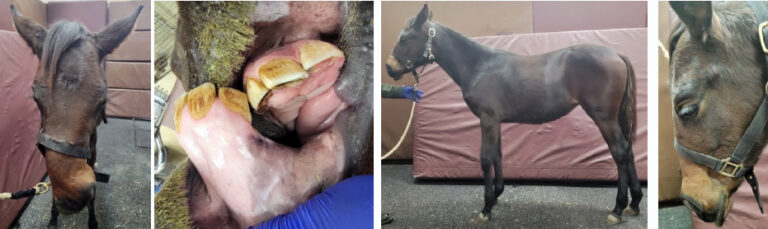
An open mouth, dorsal to ventral oblique view can isolate the crown, reserve crown, and crestal bone of mandibular cheek teeth.
“In practice this can be an important technique when imaging an older horse with a fractured tooth,” advised McAndrews.
Mandibular 11s can be difficult to image due to summation of the thick masseter muscle in the traditional ventrodorsal oblique view. McAndrews recommended taking an additional view of the mandibular 11s by directing the generator dorsal to ventral oblique about 30 degrees and caudal to cranial about 15 degrees.
“Dental radiographs are very technique-sensitive. If you aren’t happy with your image, try changing the degree of your generator by a couple of degrees at a time,” said McAndrews.
Related Reading
- Equine Tooth Extractions: Know When to Refer or Abort
- Equine Dental Repulsion Using Small Diameter Repulsion Pins
- Disease Du Jour: EOTRH
Stay in the know! Sign up for EquiManagement’s FREE weekly newsletters to get the latest equine research, disease alerts, and vet practice updates delivered straight to your inbox.