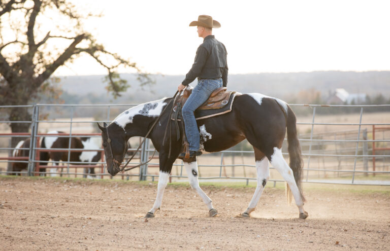
Your client calls with a “wobbly horse,” and your mind starts filtering through the list of differentials in your head. Of course, many of us can’t help but begin catastrophizing, wondering if it’s a case of equine herpesvirus myeloencephalopathy (EHM) or something not quite as dramatic but equally problematic as equine protozoal myelitis (EPM). While you’re considering these and other infectious neurologic conditions, don’t forget the small but important list of noninfectious neurologic conditions affecting horses.
The American Association of Equine Practitioners has a diagnostic flow chart available to help practitioners navigate through some of the main differential diagnoses for neurologic disease (aaep.org/resource/neurologic-disease-differential-and-diagnostic-flow-chart). In it, the “noninfectious” causes of neurologic disease include:
- Cauda equina syndrome or polyneuritis equi, the latter of which is a disease that appears as cauda equina syndrome either with or without cranial nerve abnormalities (Melvin et al. 2023).
- Temporohyoid osteoarthropathy.
- Equine neuroaxonal dystrophy (eNAD)/equine degenerative myeloencephalopathy (EDM).
- Cervical vertebral stenotic (or compressive) myelopathy (CVSM) or “wobbler syndrome,” which might include osteoarthritis (OA) of the articular facets.
- Trauma.
Of these, CVSM and eNAD/EDM are the most common causes of noninfectious neurologic disease in horses, and they are virtually indistinguishable from afar.
In this article, we’ll help practitioners recognize a noninfectious neurologic disease and describe the key features of CVSM and eNAD/EDM. We’ll also present diagnostics and treatment options, including information on rehabilitation, and briefly describe transposition of ventral lamina from C6 to C7.
First, a Complete Physical and Neurologic Examination
In severely neurologic horses, it’s typically easy to identify ataxia based on standard physical and neurologic examinations. However, mild ataxia in horses presenting with “poor performance” can certainly prove more difficult, and it can be remarkably challenging to differentiate between lameness and ataxia in some horses.
“Noninfectious neurologic diseases tend to be more chronic than infectious neurologic diseases,” explains Amy Johnson, DVM, Dipl. ACVIM (LAIM and Neurology), an assistant professor of large animal medicine and neurology at the University of Pennsylvania’s School of Veterinary Medicine. “A fever certainly suggests an infectious cause, but absence of fever does not rule out an infectious cause.”

She says clues for diagnosing EHM include urinary incontinence, vasculitis (limb swelling), rapidly progressive disease (hind end usually affected first), a history of horses moving in and out of the barn, recent travel, respiratory disease, fever, nasal discharge, and abortions. While EPM can truly mimic anything and cause either symmetric or asymmetric signs, the presence of asymmetric ataxia plus focal muscle atrophy or multifocal disease might point practitioners toward EPM.
“In contrast, both CVSM and eNAD/EDM tend to cause symmetric signs that are consistent with cervical or diffuse spinal cord disease,” says Johnson.
Infectious diseases more commonly cause brain plus spinal cord disease or even multifocal signs.
“A complete blood cell count and internal organ function test rarely help differentiate between infections and noninfectious neurologic disease unless the horse has something weird, such as a bacterial meningitis or discospondylitis,” says Johnson. “Instead, a complete neurologic examination and cerebrospinal fluid (CSF) cytology followed by specific disease testing are the most helpful tools to rule out infectious diseases.”
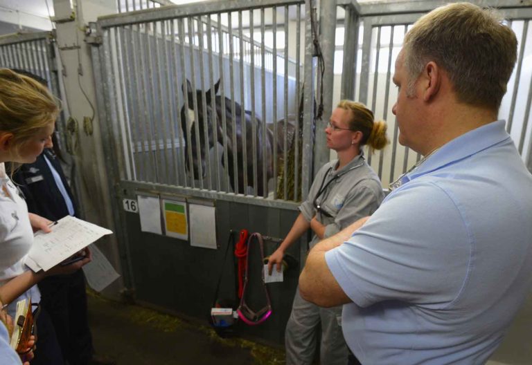
To conduct a full and complete neurologic examination, Johnson recommends starting with the horse in the stall and observing its mental status and behavior. Begin by examining the spinal reflexes, specifically the cervicofacial, cutaneous trunci, and perineal/anal reflexes as well as tail tone. While the horse is standing quietly, evaluate his posture, head and neck carriage, musculature (underdevelopment/atrophy), and areas of abnormal sensation/sweating.
During the dynamic phase of the examination, evaluate the horse’s gait. Horses must be evaluated for signs of incoordination or weakness at the walk and trot, in a straight line and a circle. Serpentine movements, walking with the head elevated, pulling the tail while the horse is walking, and tight circles will help identify ataxia, as well as walking backward, uphill, and downhill with the head in a normal position and elevated.
When evaluating neck pain, practitioners can hold a carrot at the horse’s flank to see if he will reach back toward it. Using these baited stretches, however, might give false information.
“Many horses are so food motivated they will find a way to reach their flank in a very inappropriate way,” explains Melinda Story, DVM, Dipl. ACVS, ACVSMR, an assistant professor of equine sports medicine and rehabilitation at Colorado State University’s College of Veterinary Medicine and Biomedical Sciences, in Fort Collins. “I think this is commonly misinterpreted as normal when frequently the horse is painful and dysfunctional.”
“Finish the exam by observing the horse grazing to look at neck and limb placement,” advises Johnson.
When whittling away at the long list of potential causes for the observed or reported neurologic abnormalities, Johnson reminds practitioners there are no hard and fast rules in neurology!
“EPM is often asymmetric, but it can be symmetric, whereas eNAD/EDM is usually symmetric,” she says. “CVSM can be asymmetric but is more commonly symmetric. Further, EPM can cause focal severe muscle atrophy, but it doesn’t have to. CVSM can cause neck muscle atrophy, and horses with eNAD/EDM often have topline (epaxial) and gluteal muscle atrophy that is mild to moderate (but not severe) and symmetric.”
Once you strongly suspect a noninfectious neurologic disease, the top two conditions to consider are CVSM and eNAd/EDM. CVSM occurs in approximately 1.3% of all horses, primarily in Thoroughbreds, Quarter Horses, and warmbloods.
Recognizing, Diagnosing, and Treating CVSM

CVSM is caused by spinal cord compression in the neck region due to structural abnormalities of the cervical vertebrae, joint instability and OA, or soft tissue or bony changes in the neck. Clinical signs of CVSM include incoordination and stiffness that can be more obvious in the hind limbs than the forelimbs. The horse might have abnormal head and neck posture.
Practitioners typically start by taking plain X-rays of the neck from the first cervical vertebra (C1) through to and including the first thoracic vertebra (T1). They might also obtain lateral images and oblique views.
“Classic abnormalities appreciated in horses with CVSM on these plain X-rays include malalignment (subluxation) of the joints, OA or bony proliferation, osteochondral fragments in joints, stenosis of the vertebral canal, overriding dorsal lamina, end plate flare, malarticulation, and cystic changes in articular processes,” says Johnson.
Even with the advent of large-bore magnetic resonance imaging (MRI) units, Johnson says MRIs cannot image the entire neck.
“The usual diagnostic sequence for diagnosing CVSM is survey radiographs followed by a computed tomography (CT) myelogram. Ultrasound is not helpful for diagnosing spinal cord compression,” she says.
Treating CVSM surgically involves fusion of the neck bones to eliminate movement at the affected joint spaces. One of the inaugural surgical techniques used a cylindrical implant placed in the body of two adjacent vertebrae (the “basket surgery”). But according to Johnson and Story, newer implants/techniques are available, and the surgical technique of choice is highly institution dependent. Regardless of the surgical technique, improvement in neurologic signs is likely due to remodeling of the soft tissue and bony structures in that region causing enlargement of the spinal canal.
Alternatively, CVSM can be managed medically, which is more common than surgery. Medical management includes rest/decreased exercise, anti-inflammatory drugs, and dietary modifications in young patients.
“A medical approach, however, does not remove the underlying cause and therefore is not usually curative,” says Johnson. “Further, there is no specific protocol for anti-inflammatory treatment. Some practitioners use either systemic non-steroidal anti-inflammatory drugs or corticosteroids, and others use targeted joint or perineural injections with corticosteroids or even biologic products. Medical treatment is tailored to the individual horse and lesions, meaning there is no generic recommendation.”
Medical management alone is unlikely to improve the ataxia long term, and the condition will be progressive, especially if related to OA. Surgical stabilization reportedly results in an 80% improvement in ataxia, and about 63% of horses return to athletic function. But those horses will be unlikely to compete at the same level as before surgery and are unlikely to progress in their training.
Story reminds practitioners that rehabilitation is a critical component to improve strength and neuromuscular control in any horse with neurologic dysfunction.
“The overarching goal of rehabilitation in the CVSM setting is to get the nervous system communicating correctly to the muscles for better body control,” she says. “Some horses may even become ridable, but some owners consider a case successfully managed as long as the horse is happy and safe to itself. Surgical techniques and rehabilitation programs continue to improve, and we can therefore expect outcomes associated with CVSM to also improve.”
Recognizing, Diagnosing, and Treating eNAD/EDM

eNAD/EDM is an inherited neurodegenerative disease of the brainstem and spinal cord in young horses with a vitamin E deficiency. Affected horses present with a progressive, symmetrical ataxia, paresis, and forelimb hypermetria. In some cases, the ataxia is more evident in the hind limbs, as seen in CVSM. Clinically, eNAD and EDM are indistinguishable and, therefore, typically lumped together because they are believed to be related. eNAD is considered a localized form of EDM, while EDM a more advanced form of disease. This condition can present as young as six months, but some horses do not become clinical until later in life, around 5 to 10 years of age.
Previous research suggested veterinarians could reach an antemortem diagnosis by measuring a protein called phosphorylated neurofilament heavy chain (pNfH) in serum and cerebrospinal fluid.
“This test is no longer available as it did not prove useful in eNAD/EDM setting, leaving no antemortem tests available for this condition,” says Johnson.
No genetic testing has proven interesting so far despite the strong familial suspicion.
We also don’t have a targeted treatment for horses suspected of having eNAD/EDM.
“We can’t design a therapeutic plan to a problem we can’t confirm,” says Story. “All we can do is support horse we suspect have eNAD/EDM with strengthening and mobility exercises as well as nutritional support for their nervous system.”
Preventing this disease centers on ensuring pregnant mares and foals have access to adequate vitamin E.
“I recommend testing individual horses’ vitamin E levels to determine if they need vitamin E supplementation and how much,” says Johnson. “There are no blanket recommendations for how much vitamin E these horses need, but horses at risk for neurodegenerative disease seem to need higher levels of supplementation to maintain adequate blood levels.”
Don’t Be Fooled by Transposition of Ventral Lamina From C6 to C7
Uni- or bilateral transposition of the ventral lamina from C6 to C7 (TC67) causes neck pain but not neurologic disease, says Story.
“This is a hot topic right now because it has become more commonly recognized, but we really do not know the clinical significance,” she explains. “There is also a large variability in the transposition, such as bilateral, unilateral, partial, etc. So even within the diagnosis, there is variability. And, the studies that have been published so far are very inconsistent in the findings.”
In her 2024 study, Story and colleagues reviewed medical records from 135 horses that had lateral-lateral cervical vertebral radiographs performed at Colorado State University from August 2020 to August 2022.
“In total, 27 (20%) of the horses had TC67. No significant difference in neurologic disease, lameness, or cervical orthopedic issues were identified when comparing horses with and without TC67,” relays Story.
But TC67 does appear to be associated with neck pain in this population of horses.
“Sixteen horses had neck pain, and five (31%) had TC67 whereas 11 (69%) did not. But for the horses without neck pain, 22 (18%) had TC67 and 97 (82%) did not,” Story adds.
In her opinion, X-rays are great for giving baseline information, but caution should be used to avoid overinterpretation not only of TC67 but other lesions as well, including compressive lesions and the significance of cervical articular process joint enlargement interpreted as OA.
“As advances and availability of CT imaging increases, so too will our knowledge and understanding,” Story says.
Take-Home Message
Although much less common than infectious neurologic diseases, keep CVSM and eNAD/EDM in mind when the physical, neurologic, and clinicopathologic examinations suggest a noninfectious condition. In general, the tipoffs for a noninfectious neurologic condition include chronicity and symmetrical signs consistent with either cervical or diffuse spinal cord disease.
Related Reading
- AAEP Health Coverage: Vitamin E Response for eNAD and EDM
- Cervical Ventral Laminae Transposition and Neck Pain in Horses
- Managing Horses With Low Serum Vitamin E
Stay in the know! Sign up for EquiManagement’s FREE weekly newsletters to get the latest equine research, disease alerts, and vet practice updates delivered straight to your inbox.

![[Aggregator] Downloaded image for imported item #18375](https://s3.amazonaws.com/wp-s3-equimanagement.com/wp-content/uploads/2025/09/30140031/EDCC-Unbranded-26-scaled-1-768x512.jpeg)
![[Aggregator] Downloaded image for imported item #18890](https://s3.amazonaws.com/wp-s3-equimanagement.com/wp-content/uploads/2025/11/17113845/EDCC-Unbranded-3-scaled-1-768x512.jpeg)

