Equine grass sickness (EGS) is a devastating disease with an 80% mortality rate. It can occur in both wild and domesticated equids in any temperate part of the world. The disease causes severe damage to the autonomic nervous system (which controls involuntary functions such as sweating, heart rate, and gut movement), leading to liver damage, heart damage, and total GI stasis. The development of this disease is extremely rapid, with acute cases having 100% mortality and death occurring within two days of symptoms developing. Chronic cases (lasting more than seven days) can have less severe clinical signs and approximately 50% survival rate.1 However, even these horses can have permanent nervous system damage resulting in long-term symptoms and reduced welfare.
Despite knowledge of this disease existing for more than 100 years and being reported in many countries across the globe, we still know very little about the cause. There also remains a misconception that EGS only occurs in Scotland, despite cases being reported in England, Wales, Sweden, Italy, France, Germany, Netherlands, Eastern Europe, and North and South America.
Why Are We Talking About EGS?
As we expand our understanding and research of this disease, we are realizing it’s more widespread than initially thought. We have received case reports of EGS in the United States and strongly suspect it occurs here. This isn’t meant to cause panic, however. If EGS exists in the U.S., then it likely has for a long time and is simply being misdiagnosed or underreported. Prevalence also varies year to year, and case reports remain relatively low, so the disease is not likely common. Our goal is to raise awareness among veterinarians and better understand the epidemiology of this disease.
Essentially, we’re asking you to keep EGS in the back of your mind the next time you’re faced with an unresponsive colic with some unusual clinical signs.
What Are the Risk Factors?
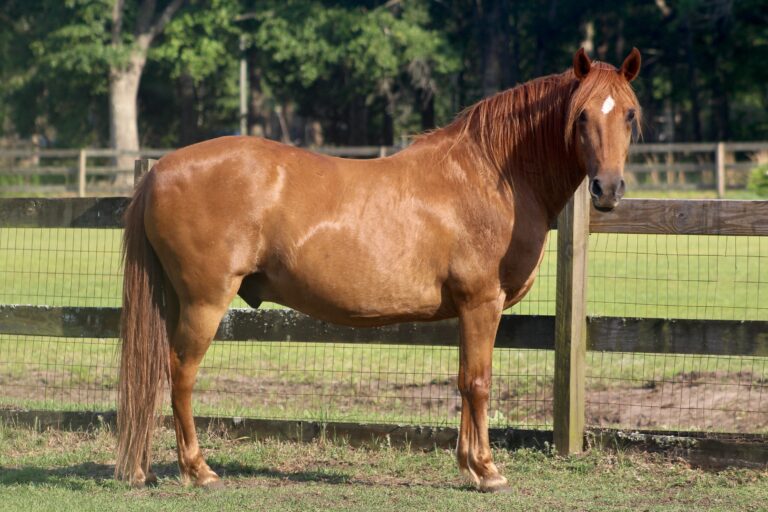
Equine grass sickness was first identified in 1907 and dubbed “grass sickness” because it is almost exclusively seen in horses eating fresh, green grass in pasture. It is not associated with stabled horses.
The biggest risk factor we’ve identified is, unfortunately, beyond our control: the season. In the UK, EGS observes its peak caseload in March through May with rising temperatures and grass growth, with a second peak in September and October. Similarly, high-risk weather events such as drought or heavy rainfall causing changes in grass growth rates and metabolism are associated with EGS cases.2
Other risk factors include changing an animal’s pasture, changes in diet and deworming strategies, and stressful events (such as travel or management changes). Disruption of the pasture or soil (induced by close grazing or construction practices) is also associated with an increased risk of EGS.2,3
Finally, the age of the horse is a strong risk factor, with younger horses between the ages of 2 and 7 being at the highest risk.
Signs of EGS

EGS is often mistaken for colic because the main sign of acute EGS is intestinal paralysis, with loss of all gut sounds. Afflicted animals might show behavioral changes such as pawing at the ground, a “tucked-up” posture, and excessive salivation. Chronic cases might also produce foul-smelling nasal discharge and display inflammation of the nasal passage. The stomach is likely to become distended due to severe constipation, and any feces passed appear dense and coated in mucus. Many horses show difficulty swallowing and food avoidance.
Due to the disease’s neurologic nature, affected horses might develop arrythmia or tachycardia (increased heart rate) alongside patchy sweating, muscle tremors, and ptosis (drooping of the eyelid).

Diagnosing EGS
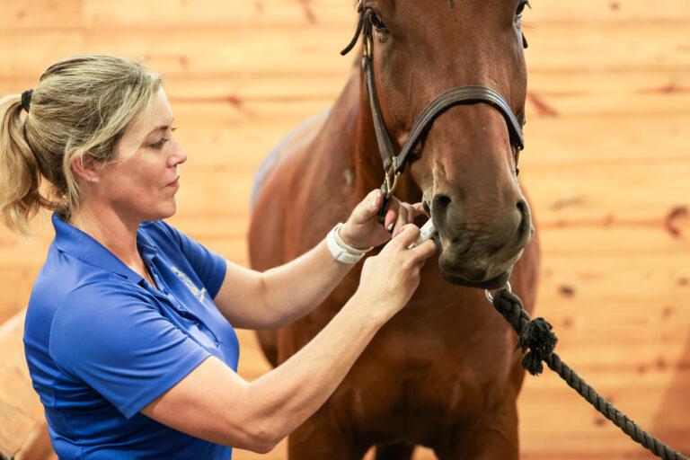
Aside from differential diagnosis and finding colic unresponsive to treatment, diagnosing EGS is difficult because we don’t yet have a straightforward, noninvasive antemortem test. Administering phenylephrine eyedrops to horses displaying ptosis can be an indicator of the neuronal damage associated with EGS.4 In the presence of EGS, these eyedrops will cause the angle of the horse’s eyelashes to change within approximately 30 minutes.
A biopsy from the ileum and rectal tissue can also be used but requires a pathologist skilled in measuring neurodegenerative changes through H&E staining. By far the easiest method of diagnosis is post-mortem sampling of the ileum and cranial cervical ganglia. Equine grass sickness causes marked degeneration of the neurons in both the ganglia and the mesenteric nervous system layer of the ileum.5
What Can You Do?
The Moredun Research Institute and the Equine Grass Sickness Fund (EGSF) has initiated the EGS Biobank project, generously funded by the British Horse Society, to collect samples from EGS cases in the UK. They also collect information about EGS cases from around the globe, which has been essential in understanding common factors across cases.
The EGSF is currently working with Equine Infectious Disease Surveillance (EIDS) to develop a real-time, international case-reporting platform that when complete will be part of the International Collation Centre. In the meantime, we’d be grateful for reports of any EGS cases through the Equine Grass Sickness Biobank Owner Questionnaire, available on the EGSF website. These reports can be made anonymously and only require as much information as you are prepared to give.
For more information or discussion around EGS and contributing to the biobank, email Dr. Beth Wells (Beth.Wells@moredun.ac.uk) or Dr. Tanith Harte (Tanith.Harte@moredun.ac.uk).
References
1. Halliwell, L. and Carslake, H. (2022), Clinical update on equine grass sickness. In Practice, 44: 476-482.
2. Wylie, C. E., Shaw, D. J., Fordyce, F. M., Lilly, A., & McGorum, B. C. (2014). Equine grass sickness in Scotland: a case-control study of signalment- and meteorology-related risk factors. Equine veterinary journal, 46(1), 64–71.
3. Wylie, C.E., Shaw, D.J., Fordyce, F.M., Lilly, A., Pirie, R.S. and MCGorum, B.C. (2016), Equine grass sickness in Scotland: A case-control study of environmental geochemical risk factors. Equine Vet J, 48: 779-785.
4. Hahn, C. N., & Mayhew, I. G. (2000). Phenylephrine eyedrops as a diagnostic test in equine grass sickness. The Veterinary record, 147(21), 603–606.
5. Piccinelli C, Jago R, Milne E. Ganglion Cytology: A Novel Rapid Method for the Diagnosis of Equine Dysautonomia. Veterinary Pathology. 2019;56(2):244-247. doi:10.1177/0300985818806051


