Equine myopathies can manifest in many ways, from classic signs of tying-up to more ambiguous aspects of poor performance. During a Burst session at the 2025 American Association of Equine Practitioners, Sarah F. Colmer, VMD, ACVIM, a large animal internist and neurology fellow at the University of Pennsylvania School of Veterinary Medicine’s New Bolton Center, reviewed common myopathies and how to diagnose and manage them, including tips for taking muscle biopsies.
Sporadic Exertional Rhabdomyolysis
Sporadic exertional rhabdomyolysis is believed to occur secondary to external factors such as overexertion, exhaustion, and poor nutrition, Colmer explained. Clinical signs (e.g., short and stilted gait, reluctance to ambulate/recumbency, firm muscles, myoglobinuria) are typically acute and develop following exercise.
The easiest way to identify cases is by measuring AST and CK levels, which can remain elevated for several weeks after an episode, she said. Treatment is variable but includes analgesia, hydration, NSAIDs, rehydration with electrolyte support, rest (sometimes several weeks), and diet alterations.
Recurrent Exertional Rhabdomyolysis
In contrast to sporadic cases, said Colmer, these horses are suspected to have intrinsic muscle dysfunction, and signs can occur with minimal exercise.
“CK and AST may be normal at rest, making it harder to diagnose these cases,” she added.
She suggested veterinarians in the field consider doing exercise response tests on horses they suspect have recurrent exertional rhabdomyolysis. This involves obtaining baseline CK and AST values prior to exercise and 4-6 hourspost-exercise. A positive test is marked by significant elevation (3-4X increase) in CK and AST. “Fifteen minutes of trotting on a longe is sufficient to produce this response,” said Colmer.
Management, however, is quite extensive and involves trying to minimize stress, keeping affected horses calm and comfortable, and addressing clinical signs as they become apparent.
Polysaccharide Storage Myopathy
Clinical signs of PSSM include stiffness, soreness, muscle fasciculations, sweating, and more subtle signs of poor performance as well as lameness. Veterinarians can diagnose PSSM1 cases using a hair sample, whereas PSSM2 cases can only be diagnosed on muscle biopsy, said Colmer.
“Management is relatively extensive, but around 70% of horses can improve,” she said, recommending regular, consistent exercise for these horses with as few days off as possible and working with an equine nutritionist to reduce starch and sugar intake.
Myofibrillar Myopathy
Colmer described this disease as rooted in a disarray and ectopic accumulation of the cytoskeleton protein desmin at a molecular level, which Dr. Stephanie Valberg has identified and has been researching at Michigan State University.
“Arabians have their own special flavor (MFM-ER) in which they have more of an exertional rhabdomyolysis presentation and increased CK and AST,” she explained. “But our warmbloods with MFM can present with insidious exercise intolerance around the age of 6-8 years.”
The latter population might have difficulty with collection, cantering, lack of impulsion, stiffness, and reluctance behaviors such as bucking and rearing. “So when we’re evaluating these horses and our owners mention very specifically that they have a reluctance to move forward when they apply leg pressure or they have this sort of exercise intolerance that develops over time, our ears kind of prick, and we consider the possibility of MFM,” she said.
Veterinarians use biopsy to confirm a diagnosis. Colmer recommends dietary management for these horses in collaboration with an equine nutritionist. They require moderate NSC and low fat intake, high levels of protein (12%), and possibly supplementation with co-enzyme Q10, N-acetylcysteine (MFM Pellet), and vitamin E.
Arabians with MFM-ER require daily exercise, while warmbloods seem to do better with days off (e.g., three days on, two days off), she said. They benefit from long warm-ups, long and low work, and core strengthening.
“A lot of horses when management is pursued will improve within four weeks,” said Colmer. “Around 71% of warmbloods improved in one study, but we have less data on Arabians.”
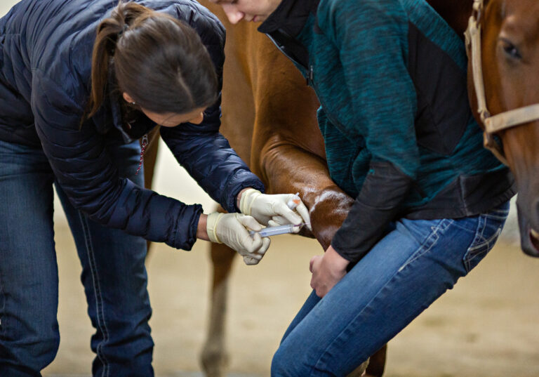
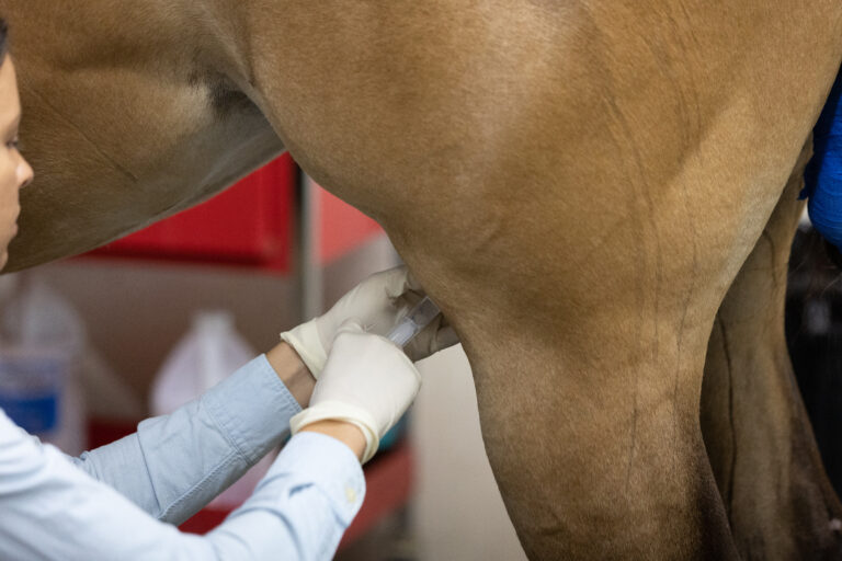
Muscle Biopsies
Lastly, Colmer demonstrated the Bergstrom biopsy technique, which she said has revolutionized the way she takes biopsies. It’s simple, safe, much faster, and less invasive than the traditional semimembranosus/semitendinosus, or incisional, technique.
Using a Bergstrom biopsy instrument, you make a small incision in the skin and subcutaneous tissue over the horse’s middle gluteal muscle. Then, insert the biopsy instrument about 3 inches deep. Move the inner piece of the instrument up and down around 10-12 times to produce a “sausage” of muscle within the instrument that you can then remove and submit for testing.
Take-Home Message
Myopathies have many manifestations, some of which can be insidious and present as performance issues, said Colmer, adding that she hoped attendees come away with confidence to pursue muscle biopsies.
Related Reading
- Demystifying Equine Muscle Disease
- Diagnosing Performance-Limiting Muscle Diseases in Horses
- Type 2 PSSM in Quarter-Horse-Related Breeds
Stay in the know! Sign up for EquiManagement’s FREE weekly newsletters to get the latest equine research, disease alerts, and vet practice updates delivered straight to your inbox.