Equine Dental Repulsion Using Small Diameter Repulsion Pins 

A retrospective case series described the outcome for 20 cases after dental repulsion using small diameter repulsion pins. 
Pre-operative lateral (left) and 15° ventro-dorsal lateral oblique radiographs of the same case showing crosshatch staple placement to facilitate accurate positioning of trephine hole below of the affected 309.
Pre-operative lateral (left) and 15° ventro-dorsal lateral oblique radiographs of the same case showing crosshatch staple placement to facilitate accurate positioning of trephine hole below of the affected 309. | Kamps M, Barakzai S. Equine Vet J. 2024.

Reported complication rates after dental repulsion for equine exodontia are high (up to 80%), but repulsion methods have changed notably in the last 20 years. The aim of this retrospective case series was to describe the outcome for 20 cases after dental repulsion using small diameter repulsion pins. 

Inclusion Criteria

Researchers reviewed clinical records of horses that underwent cheek tooth repulsion (2014-2023). Inclusion criteria were: mandibular or maxillary cheek tooth extraction where oral extraction failed and repulsion was used to complete extraction and where clinical follow-up information was available. Repulsions were carried out under sedation with a regional nerve block or under a short general anesthetic using a small diameter repulsion pin (3-5 mm). Intraoperative radiographs facilitated instrument placement. The alveolus was packed with polymethyl methacrylate post-extraction. Horses were reexamined four to six weeks postoperatively. 

Results
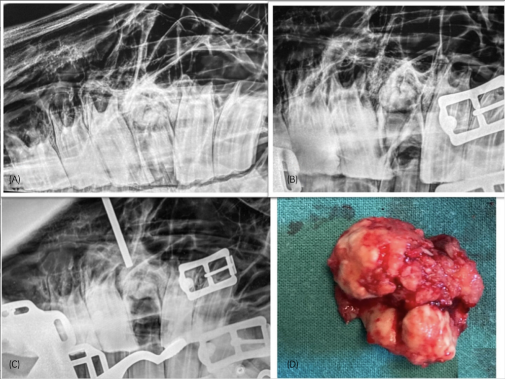
(A) Radiograph taken prior to attemped oral extraction, showing a grossly demineralized (radiolucent) 209, with radio dense apical hypercementosis and chronic midline sagittal fracture. The 208 and 210 have both drifted into the fracture site. (B) After failed oral extraction—the tooth fractured into multiple pieces—with dental material lining the alveolus and still present at the apical area. The apical fragment was firmly attached and could not be loosened with dental picks. (C) An intra-operative radiograph confirming 5 mm pin placement over the apex, prior to repulsion. (D) Apical hypercementosis—after repulsion.
(A) Radiograph taken prior to attemped oral extraction, showing a grossly demineralized (radiolucent) 209, with radio dense apical hypercementosis and chronic midline sagittal fracture. The 208 and 210 have both drifted into the fracture site. (B) After failed oral extraction—the tooth fractured into multiple pieces—with dental material lining the alveolus and still present at the apical area. The apical fragment was firmly attached and could not be loosened with dental picks. (C) An intra-operative radiograph confirming 5 mm pin placement over the apex, prior to repulsion. (D) Apical hypercementosis—after repulsion. | Kamps M, Barakzai S. Equine Vet J. 2024.

The study included 20 cases. Patients had a mean age of 10.3 years (range 5-16 years). The majority (75%) of teeth had preexisting dental fractures. Maxillary (n = 15) and mandibular cheek teeth (n = 5) were all successfully repulsed, with 16 cases performed with the horse standing and four with the horse under general anesthesia. Intraoperative complications included damage to the mandibular bone (n = 1). Short-term complications (n = 2) included superficial surgical site infection and dehiscence of one sinus flap. Long-term complications included the recurrence of sinusitis (n = 1) and small intra-alveolar fragments causing persistent bitting problems in another patient. 

Bottom Line 

When oral extraction fails, cheek tooth repulsion using small diameter repulsion pins is an effective extraction technique. The total intra- and postoperative complication rate was 25%. 

https://beva.onlinelibrary.wiley.com/doi/10.1111/evj.14116

Stay in the know! Sign up for EquiManagement’s FREE weekly newsletters to get the latest equine research, disease alerts, and vet practice updates delivered straight to your inbox.

categories
tags
Trending Articles
Purina Farm 7
Managing Horses With Low Serum Vitamin E 
Purina Farm 5 – Andrea Brueggemann
Feeding Horses That Can't or Won't Eat
Showjumping of horse and rider jumping
Disease Du Jour: Role of the Permitted Treating Veterinarian 
Female Vet Examining Horse In Field With Owner
Veterinary Wellness Briefs: Setting Boundaries with Clients in Equine Practice 
Newsletter
Get the best from EquiManagement delivered straight to your inbox once a week! Topics include horse care, disease alerts, and vet practitioner updates.

"*" indicates required fields

Name*
Country*

Additional Offers

Untitled
This field is for validation purposes and should be left unchanged.