Equine standing positron emission tomography (PET) has been a game changer for monitoring racehorse health and guiding training and medical care, which has contributed to reduced injuries and fatalities. However, PET is not just for racehorses and can be beneficial for all horses.
With the rapid expansion in the availability of this technology, PET is becoming more accessible to diverse populations of horses. Standing PET can be used from the foot to the knee in the front limbs and the foot to the hock in the hind limbs. As a functional imaging technique that detects biological changes in processes such as metabolism and blood flow, it has the ability to “see” injuries to bone and soft tissues at the molecular level, before changes would otherwise be apparent.
Pioneered at UC Davis in 2015, equine PET imaging initially required horses to undergo general anesthesia. Modifications to allow PET imaging on standing horses under sedation greatly increased its applications and expanded its reach. Scanners have been installed at several racetracks and veterinary hospitals in recent years. Ten different sites are expected to be equipped with the technology nationwide by the end of 2023.
PET imaging has become simple with the equine standing MILEPET scanner. A small dose of a radioactive dye is injected intravenously about 30 minutes prior to imaging. This dye distributes through the body and accumulates at sites of active injuries. The horse is then sedated similarly to other veterinary procedures. The scanner is wheeled up to the horse, and an openable ring of detectors loosely closes around the limb. It only takes three to five minutes to image each site of interest. In twenty minutes, both front feet and fetlocks can be imaged with PET.
To date, more than 300 horses have received PET scans at the UC Davis veterinary hospital. Cases that have benefited from this advanced imaging modality include navicular bone injury, laminitis and joint disease. Equine patients have included American Quarter Horses, warmbloods, Thoroughbreds, and a mule. Since scans under standing sedation are more accessible and economical than those that require general anesthesia, they can easily be repeated to monitor healing, optimize treatment and inform rehabilitation for the best outcomes.
Different imaging modalities can also be combined to provide the most accurate picture. In some cases, PET and X-rays will provide all the information necessary to decide on a treatment plan. In other cases, MRI of a specific area can be performed after PET. The combination of PET and MRI data provides the most comprehensive assessment of the situation.
The use of PET will keep growing in the sport and pleasure horse populations. Ease of use and affordability make it an excellent first choice for advanced imaging. In addition to identifying injuries, the “functional” imaging properties of PET—assessing the activity of injuries—are particularly helpful for monitoring rehabilitation.
Case #1: Jool the Mule

A mule named Jool was the first equine imaged with the MILEPET scanner when it was installed at the UC Davis veterinary hospital. A well-known patient at UC Davis, Jool had been suffering from lameness due to chronic tendon lesions that initially improved with the use of stem cell therapy but later recurred. The goal of the PET scan was to assess whether the tendon was still the main issue or if other injuries had developed. The PET scan demonstrated that, in addition to the tendon lesion, the navicular bone was also part of the trouble, which was not apparent on an MRI. This finding led to modifying Jool’s shoeing in order to protect her navicular bone.

Images from Jool’s foot. The top row are MRI images; the bottom row are PET images overlaid on the MRI images. The bright yellow spot highlights a bone injury in the navicular bone that was not recognized on the MRI.
Case #2: Hock Lameness

Another example of a case scanned with the MILEPET at UC Davis was an American Quarter Horse with lameness localized to the hock but no definitive answers on X-rays and ultrasound. This mare was the first to have standing scans of the hock performed with the MILEPET. The scans were successful and demonstrated early joint disease that was responsible for the pain. The PET results helped target the exact area of the joint to inject.
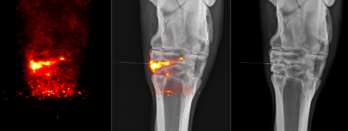
The image to the left is one of the obtained PET images. The yellow area indicates abnormal bone. When overlaid with the X-ray (middle image), it shows that the injury is centered on one of the small joints of the hock, despite minimal changes on the X-ray (image on the right). With this information, the abnormal joint was injected with corticosteroids to alleviate the pain.
This article originally appeared in the UC Davis Spring 2023 Horse Report: https://cehhorsereport.vetmed.ucdavis.edu/news/positron-emission-tomography-pet.