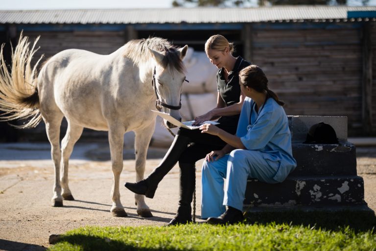
Horses perform in a variety of athletic endeavors that depend on different skill sets, but one thing they have in common is the development of lameness issues.
Equine veterinary medicine often leads the way in managing degenerative arthritis, using strategies now being employed for human patients. What new developments have been in the works for lameness issues in horses?
Gene Therapy Using AAVIRAP
Clinicians and researchers at the Orthopaedic Research Center at Colorado State University have investigated methods to reduce inflammation over a long period using gene vectors, specifically adenoassociated viral vectors (AAV) with the DNA sequence that encodes for interleukin receptor antagonist protein (IRAP). The objective is to maximize protein production in a joint to assist cartilage and bone repair, which have “extended healing times,” while not inducing further inflammation within the joint.
IRAP works its anti-arthritic effects by blocking or antagonizing interleukin- 1, a cytokine present in degenerative arthritic joints. A recent study injected AAVIRAP into the middle carpal and metacarpal-phalangeal joints of two horses, which resulted in good success in elevating IRAP levels for at least four months. The injections did not elicit any toxic effects, swelling or pain in the injected joints. The goal of providing longer-term IRAP anti- inflammatory activity was achieved in this trial, and the horses’ progress continues to be monitored.
Muscle Studies
Myofibrillar myopathy (MFM) is undergoing research at the Michigan State University’s Equine Neuromuscular Diagnostic Laboratory by Stephanie Valberg, DVM, PhD, DACVIM, ACVSMR, and colleagues. This newly identified syndrome involves the disruption of the orderly alignment of myofibril contractile proteins.
The test to confirm MFM relies on staining desmin in a muscle biopsy rather than on genetic testing. According to reports from the Neuromuscular Diagnostic Laboratory, “Normally desmin is found at one specific repeating place in the myofibril, the Z-disc, and it acts to hold myofibrils in proper alignment across the muscle cell. In horses with MFM, sections of a muscle cell start to produce abnormal amounts and shapes of desmin, likely as a reaction to instability in the contractile proteins.”
Glycogen might pool in the breaks between myofibrils, thereby eliciting a false positive diagnosis of Type 2 equine polysaccharide storage myopathy (PSSM2). Instead, Valberg and colleagues note that MFM is a separate pathology of muscle. MFM might be incorrectly diagnosed if a biopsy is taken from a horse with actively regenerating muscle tissue following an injury.
Warmblood horses: Researchers have identified that myofibrillar myopathy, or MFM, occurs in some warmblood horses (Valberg, S.J., et al. Clinical and histopathological features of myofibrillar myopathy in warmblood horses. Equine Vet J 2017 May 22). Muscle disease can significantly interfere with a horse’s athletic function. Some warmblood horses by ages 8-10 might begin to experience exercise intolerance, reluctance to go forward, stiffness, and hind limb lameness that is not readily localized through diagnostic procedures.
Data was obtained through muscle biopsies of 10 horses that experienced myopathy signs. Cellular evaluation and staining revealed “desmin-positive aggregates in myofibres of the founding dam and in horses from two subsequent generations.” The study concluded that MFM is a potentially heritable form of exertional myopathy.
Arabian horses: Intermittent episodes of exertional myopathy are not uncommon findings in Arabian horses, especially those engaged in endurance pursuits. Muscle biopsies were obtained in 14 Arabian controls, 13 Arabian horses with complaints of myopathy and 25 samples evaluated from Arabians previously classified as having Type 2 polysaccharide storage myopathy (PSSM), recurrent exertional rhabdomyolysis (RER) or no pathology (Valberg, S.J., et al. Suspected myofibrillar myopathy in Arabian horses with a history of exertional rhabdomyolysis. Equine Vet J 2016 Sept:48(5):548-556). Rather than identifying a storage myopathy, the study discovered that these horses were experiencing myofibrillar myopathy.
Management of MFM: While the predominant breeds associated with MFM are Arabians and Warmbloods to date, cases have been identified in a few Thoroughbreds, Quarter Horses and Paso Finos. Nutritional changes to providing low-starch, low-sugar diets might be helpful. Fat can be included if necessary to maintain body condition. Whey protein can be helpful to add bulk to muscle, especially when fed 45 minutes prior to exercise. However, diet alone cannot cure this syndrome.
Valberg’s group recommended avoiding rest and instead implementing a steady program of incremental and consistent training, along with pasture turnout. These horses do best with a good warm-up period that stretches the topline and core abdominal muscles via a long-and-low frame for five to 15 minutes before more intensive work is requested.
Early Exercise in Young Foals
Horse owners often perceive that young foals should be protected from too much exercise. By keeping foals in confinement, they might be doing the young horses a great disservice.
Wayne McIlwraith, BVSc, MS, PhD, DSc, FRCVS, DACVS, DECVS, DACVSMR, of Colorado State University (CSU), has pioneered many advances in the field of equine orthopedics. He collaborated with Chris Kawcak, DVM, PhD, DACVS, also of CSU, and Elwyn Firth, BVSc, MS, PhD, DACVS, DSc, of the University of Auckland, in a recent investigation into the effects of early exercise on the metacarpal-phalangeal joint (Kawcak, C.E.; McIwraith, C.W.; Firth, E. Effects of early exercise on metacarpophalangeal joints in horses. Am Vet J Res 2010;71:405-411).
Six pasture-reared horses preconditioned with exercise from 3 weeks of age to 18 months of age were compared to six similar-aged horses that were provided with only daily pasture turnout. Significant differences were identified.
“Horses that were exercised since near birth had fewer gross lesions in the joints, greater bone fraction in the dorsolateral aspect of the condyle, and higher bone formation rate compared to non-exercised horses,” noted the research. “However, there was less articular cartilage matrix staining in the dorsal aspect of the condyles in exercised horses.”
Young horses exercised at an early age appear to benefit from the protective effects of exercise on the fetlock joints; however, reasons for reduced cartilage matrix staining deserve further investigation.
A similar study had previously reviewed the effects of early exercise on mid-carpal joints and found no deleterious effects (Kim, W.; Kawcak, C.E.; McIlwraith, C.W.; Firth, E.C.; McArdle, B.; Broom, N.D. Influence of early conditioning exercise on the development of articular surface abnormalities in cartilage matrix swelling behavior in the equine middle carpal joint. Am J Vet Res 2009;70:589-598). “Investigators have concluded that early conditioning is a positive influence on articular cartilage and is safe to use.”
Injection of the Navicular Bursa in a Lateral Approach
Treatment of podotrochlear disease is a common procedure for equine practitioners. A newer technique enables access to navicular bursa injections without penetration of the deep digital flexor tendon.
A lateral approach to the navicular bursa using ultrasound guidance was performed on bilateral forelimbs of 62 cadaver horses and 26 live horses (Nottrott, K.; De Guio, C.; Khairoun, A.; Schramme, M. An ultrasound- guided, tendon-sparing, lateral approach to injection of the navicular bursa. Equine Vet Journal 2017 Jan 27). Radio-contrast agent was injected with the foot placed in a flexed position on a navicular block.
In 91% (104/114) of the limbs injected, contrast agent was identified in the navicular bursa. Contrast was deposited in only the navicular bursa and no other structures in 78% of the limbs injected. In those cases where material did not get injected into the navicular bursa, failure was attributed to poor ultrasound quality, leading to injection of the coffin joint, tissue around the bursa and, in one horse, the deep digital flexor tendon.
One caveat of this study is that the injections performed were done on non-diseased navicular bursae. It is undetermined how well this technique will work with cases of clinical disease of the podotrochlear apparatus, especially in regard to injection of diagnostic anesthesia and therapeutic agents.
Comparison of Stem Cell Aspirates From the Ilium or Sternum
Regenerative therapies are instrumental in returning horses to athletic usefulness. Much research has been directed toward the use of stem cells and, in particular, bone marrow aspirates of bone mesenchymal cells.
A study at the Colorado State University’s Equine Orthopedic Research Center compared quality of aspirates in seven horses aged 2-5 years. While no differences in cell numbers or growth rate were identified between either stem cell sources—ilium versus sternum—what was of significance is that in both sites, the first five milliliters of aspirate “yielded the highest concentration of stem cells.”
Take-Home Message
One of the attributes horse owners seek in a veterinarian is a willingness to keep up with medical knowledge. EquiManagement strives to alert equine veterinarians to research updates through the Keeping Up segment in each magazine, through additional medical and research articles on EquiManagement.com, through our monthly Research Reports newsletter and through links to articles from referred journals on EquiManagement’s Facebook page.


