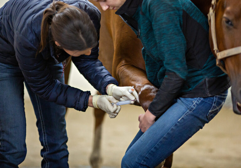
To prepare for sterile enucleation in the field, most frequently performed via the transpalpebral approach, adequately sedate the horse. Use detomidine (and maybe even morphine) and a longer-acting local anesthetic to perform a retrobulbar block (through the supraorbital fossa or via the four-point approach) using spinal needles. In addition, have a handler at the horse’s head, which should be resting on a solid structure such as hay bales, shaving bags, or a garbage can.
“The first step is a quick tarsorrhaphy followed by a fusiform skin incision close to the eyelid margin,” said Kelly Knickelbein, VMD, DACVO, Assistant Professor in the Section of Ophthalmology at the Cornell University College of Veterinary Medicine, during a Burst presentation at the 2024 AAEP Convention. “If your incision is too far from the eyelid margin, there will be too much tension on the skin when closing. But if you’re not far enough, then you won’t be removing the meibomian glands.”
Next, dissect to—but not through—the conjunctiva with Metzenbaum scissors. Use a blade to transect the medial and lateral canthal ligaments. Once these ligaments are released, the globe can rotate more freely. Then switch to curved mayo scissors to work more posteriorly until the globe can be rotated 360 degrees.
Knickelbein emphasized the importance of ensuring complete removal of all necessary tissues when enucleating horses, including the entire globe, the third eyelid and its cartilage and gland, all the conjunctiva, the eyelid margins (containing the meibomian glands), and the medial caruncle. Clamping or ligating the optic nerve is not necessary. The incision should be closed in at least two layers.
“You don’t have to place a bandage, but I like to because it keeps the incision clean and limits the amount of swelling that occurs,” said Knickelbein. “It is critical that the bandage not slip and rub the remaining eye.”
Further, providing postoperative pain management with systemic NSAIDs and administering a systemic antibiotic such as trimethoprim sulfamethoxazole is indicated. Knickelbein recommended activity restriction to a stall or small paddock for 10-14 days until suture removal.
Always submit the eye for histopathology. “You never know what you might find if you don’t look!” said Knickelbein.
Related Reading
- Successful Subpalpebral Lavage Placement in Horses
- Prepurchase Ophthalmic Examinations: What Do You Need?
- Researchers Develop Eye Drop to Treat Uveitis in Horses
Stay in the know! Sign up for EquiManagement’s FREE weekly newsletters to get the latest equine research, disease alerts, and vet practice updates delivered straight to your inbox.


