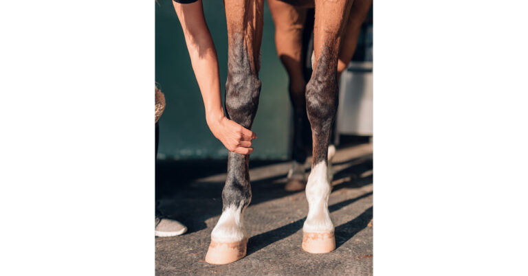
Subpalpebral lavage (SPL) systems allow for frequent application of ophthalmic solutions to the horse’s eye without the need to manipulate the eyelids. Many ocular diseases in horses cause significant pain or result in the globe being fragile; SPLs not only make treatment easier for the caretaker, but they can also improve the horse’s comfort.
SPL Placement: Upper or Lower Lid?
Which eyelid should you place it in, upper or lower?
“Whichever one you’re most comfortable with, as long as the footplate of the SPL does not contact the cornea,” said Kelly Knickelbein, VMD, DACVO, Assistant Professor in the Section of Ophthalmology at the Cornell University College of Veterinary Medicine, during a Burst presentation at the 2024 AAEP Convention. “The literature supports fewer complications with placement in the lower lid, which is why it is my preference.”
With lower lid placement, the third eyelid is a barrier that prevents the SPL footplate from contacting the cornea, so there is less risk of a footplate-induced ulcer.
SPL Placement Procedure
To prepare, have sedation, topical and injectable anesthetics, dilute betadine solution to prep the eye, white tape, suture, sterile gloves, and rubber bands for braiding the forelock and mane at the ready.
Begin by sedating the patient then performing an auriculopalpebral and frontal/supraorbital nerve block. Then inject local anesthetic subcutaneously at the SPL insertion site and sites of suture placement. Apply liberal topical anesthetic to the ocular surface, and clean the ocular surface with dilute betadine solution. Test the blocks with your finger before you start, and apply firm pressure to the site of intended SPL placement.
“If the horse doesn’t tolerate pressure from your finger, they are not going to tolerate the large, sharp trocar,” advised Knickelbein.
If the horse is intolerant, administer more sedation or place a twitch to ensure he doesn’t move with the sharp trocar next to the eye.
“After placing the lavage system, palpate the footplate location to ensure that it is deep in the conjunctival fornix and not at risk of rubbing the cornea,” said Knickelbein.
Suture to loose skin, and avoid suturing down the center of the face, as the skin adheres too tightly to the underlying bone in this area. Don’t encircle the SPL line with the sutures. Instead, place interrupted sutures on both sides of the line using a white tape butterfly.
“Finally, secure the line with the attached injection port to a tongue depressor covered by white tape, and tape the tongue depressor to a braid near the withers,” Knickelbein recommended.
Related Reading
- Prepurchase Ophthalmic Examinations: What Do You Need?
- Researchers Develop Eye Drop to Treat Uveitis in Horses
- Disease Du Jour: Equine Recurrent Uveitis
Stay in the know! Sign up for EquiManagement’s FREE weekly newsletters to get the latest equine research, disease alerts, and vet practice updates delivered straight to your inbox.