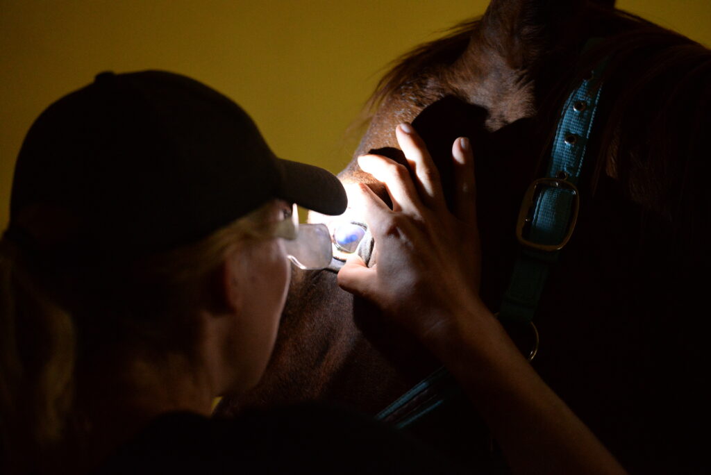
In cases where disease progresses quickly and sometimes irreversibly, time is of the essence, and the first steps taken to stabilize and address the problem matter most. Eye emergencies in horses are prime examples. Because the equine eye is known for being fragile and unforgiving, knowing how to swiftly address ocular emergencies in the field—and also when to refer cases to specialists—is an essential skill for general practitioners.
We’ve asked two experienced, board-certified ophthalmologists to share best practices with veterinarians who might be called out for equine eye emergencies on the farm and be expected to guide horse owners through the proper first-aid steps until help arrives.
Part 1: Providing Guidance to the Horse Owner
Instruct on proper first aid steps while waiting for the veterinarian.
“What are the first steps a horse owner should take following a traumatic eye injury while waiting for their vet to arrive? Take a deep breath, and remain calm,” begins Caryn Plummer, DVM, DACVO, professor of comparative ophthalmology at the University of Florida College of Veterinary Medicine, in Gainesville. “Have the owner segregate the horse from other animals and make sure there are no objects or hazards that could lead to further injury, especially if he is visually impaired. Keeping the horse calm is critical.”
No matter what caused the traumatic injury, Plummer stresses that the owner should not try to force open the eye or apply any medications until it has been evaluated. If a protective eye mask, or even a fly mask, is available, it can be used to prevent rubbing of the eye, though care must be taken that the mask is properly fitted and does not itself rub the eye. “As the veterinarian responding to the call, you may decide to recommend sedation or a systemic pain medication over the phone if you know the animal and have some understanding of his overall condition,” Plummer notes.
Explain the importance of prompt veterinary intervention.
“Delayed treatment will change the prognosis for virtually every eye emergency,” states Alison Clode, DVM, DACVO, an independent veterinary ophthalmologist consulting at Rhinebeck Equine and the New England Equine Medical and Surgical Center. For instance, an eyelid wound sutured too late will granulate and have an increased risk of dehiscence; an infected corneal ulcer diagnosed and treated too late can lead to perforation and potential enucleation; and uveitis left to progress can result in irreversible vision loss, she said.
Explaining the potential consequences of waiting to obtain a diagnosis and initiate treatment can help horse owners understand that making that call early is the safer alternative and will pay off in the long run. After initial evaluation and treatment of the eye, clients who choose to manage the horse on the farm—rather than hospitalize—should be prepared to administer oral and ophthalmic medications around the clock. In addition to almost always being emergencies, eye problems in horses are known to be time-consuming and labor-intensive, something you should emphasize to horse owners when devising a treatment plan.
Advise to hold off on applying steroidal eye ointments.
Steroids are known contraindications for corneal ulcers, but not every horse owner is aware of the risk of using them before ruling out ulceration. Plummer says it can be tempting for a client who has been repeatedly treating their horse with a topical steroid such as NeoPolyDex for an inflammatory condition (e.g., uveitis, keratitis) to just start treating an eye with that steroid when signs recur, because they assume they’re treating a flare-up of the same thing.
“This is potentially quite dangerous,” she warns. “Many types of eye disease can present with very similar signs. For example, an eye with uveitis may appear squinty, cloudy, red, and have discharge. An eye with an ulcer can have the same clinical signs. If a corneal ulcer is not identified and steroids are applied topically, that can make things much, much worse. Steroids can delay healing, increase the likelihood of infection, and even lead to complications like corneal melting. That’s why it is so very important for clients to understand the importance of having their vet out to stain the eye and make sure a corneal ulcer is not present before starting treatment with a steroid.”
Part 2: Guidance for the Practitioner
Being prepared starts with stocking the vet truck.
Clode, who travels between equine veterinary clinics to perform ophthalmology consultations and procedures, lists essential supplies for field veterinarians managing eye emergencies to carry on their truck. These include:
- Topical local anesthetic (proparacaine or tetracaine).
- Fluorescein strips.
- A focal light source, preferably with a blue (not green!) light option for visualizing fluorescein uptake.
- A direct ophthalmoscope to visualize the back portion of the eye (some have a blue light option on them) and some form of magnification (e.g., loupes).
- A tonometer to measure intraocular pressure (Tonopen or TonoVet).
- A smartphone. “Photos are extremely helpful for monitoring progression of a lesion, sharing with other colleagues who may be checking the horse, and also to send to a veterinary ophthalmologist for additional input as needed,” Clode explains.
Recognize red flags during an exam that should prompt referral to a specialist.
During an ophthalmic exam, as the attending veterinarian you will need to visually evaluate the cornea, sclera, anterior chamber, lens, iris, pupil, eyelids, and adnexa (including the eyelids and conjunctiva) and look for signs of pain, discharge, cloudiness, or trauma. You may measure the intraocular pressure and/or use a fluorescein dye to stain the cornea and look for evidence of an ulcer.
“The most common eye emergencies are trauma (eyelid/adnexal lacerations, corneal ulcers/lacerations, blunt trauma to the globe), corneal infections (infected ulcers or stromal abscesses), and acute uveitis,” says Clode. “Referral to an ophthalmologist should be considered for any adnexal laceration that is not repairable in the field, any deep corneal wound—either an infected corneal ulcer or a corneal laceration—and any eye that is acutely cloudy, blind, or uncomfortable for which an underlying diagnosis cannot be determined or managed.”
She says the most important part of managing an eye emergency is getting a clinical diagnosis. “If you can determine the clinical diagnosis, then the treatment options will follow.”
Plummer flags findings that should prompt urgent referral to an ophthalmologist:
- Large or deep ulcers.
- Penetrating wounds (including lacerations and especially foreign bodies).
- Very soft, malacic corneas where the surface contour is irregular or bulging.
“These corneal wounds can progress rapidly and often require more frequent and aggressive medical therapy than can be provided in the field,” she warns. “Also, if there are any concerns about the intraocular pressure and the field practitioner does not have access to a tonometer, referral is recommended.”
Take all corneal ulcers seriously.
Corneal ulcers are at known risk of painful rupture and subsequent vision loss in horses. “No corneal ulcer is ‘routine,’ ” says Plummer. All should be taken seriously and diagnosed and treated as soon as possible. “Many simple ones do heal quickly without complications, but even those have the potential to worsen or become infected,” she says.
“Even a superficial ulcer will be accompanied by discomfort (squinting, rubbing, tearing) and mild cloudiness,” Clode adds. Both ophthalmologists list warning signs that should raise the level of concern when evaluating a corneal ulcer:
- Marked cloudiness of the cornea (“windshield” appearance) beyond the edge of the ulcer.
- Cloudiness of the anterior chamber.
- A cream-colored or yellow appearance to the cornea, or yellowish cellular infiltration accompanying the stromal loss.
- A defect or divot (area that looks deeper than the surrounding tissue).
- A protrusion off the corneal surface.
- The ulcer is deeper than superficial. “Superficial is epithelial loss only, and the epithelium is about as thick as a piece of paper. If an ulcer looks any deeper than that, it is not superficial,” Clode warns. Histologically, the equine cornea comprises three primary layers: the superficial epithelium, central stroma, and deep endothelium.
- A miotic (small, constricted) pupil.
- Worsening of pain.
Know how to manage pain appropriately.
“Most acute eye conditions are going to be painful,” Clode says. “This pain is best treated with systemic non-steroidal anti-inflammatory drugs like flunixin meglumine, less frequently with systemic steroids, topical anti-inflammatories, topical antimicrobials, and topical atropine. In some cases, other medications such as acetaminophen, gabapentin, or trazodone can be used to help manage pain and/or behaviors that may increase risks to the globe.”
She says in her experience, the use of protective eye masks can help limit the potential for exacerbation of pain via self-trauma.
Final Thoughts
Plummer and Clode agree that, depending on the nature of the infection, in the absence of appropriate diagnosis and treatment an infected ulcer can progress to rupture within hours. All ulcers should be considered significant, and all traumatic eye injuries should be seen as emergencies warranting prompt veterinary involvement.
“It’s also important for each individual practitioner to recognize their comfort level with ocular exams, diagnoses, and treatments and to be proactive in seeking help and guidance early on in managing eye cases,” Clode says.
“It is never wrong to ask for help if you aren’t sure what you are looking at,” Plummer adds. “Veterinary medicine is a team sport!”
Researchers Explore Common Types of Ocular Conditions in Horses
In a recent retrospective study of 140 cases of clinically significant equine ocular disorders, the most commonly diagnosed primary conditions were nontraumatic keratitis (36), equine recurrent uveitis (31), traumatic injuries (22), ocular and periocular neoplasms (19), and uveitis and/or endophthalmitis resulting from sepsis (18). Nontraumatic keratitis was frequently accompanied by anterior uveitis (22), corneal rupture (16), preiridal fibrovascular membrane formation (13), and secondary mycotic infection (11).1
In a different study, researchers found that out of 446 horses with ocular disease, 61 (36%) had been diagnosed with a corneal ulcer, 44 (10%) had multiple occurrences of corneal ulcers, 65 (15%) had uveitis, and 15 (3%) had immune-mediated keratitis (IMMK).2 Blepharospasm, corneal ulceration, and uveitis can occur with IMMK, and the presence of blepharospasm and uveitis increases the odds of enucleation.3
- Flores, M. M., Piero, F. D., Habecker, P. L., & Langohr, I. M. (2020). A retrospective histologic study of 140 cases of clinically significant equine ocular disorders. Journal of Veterinary Diagnostic Investigation. https://doi.org/10.1177/1040638720912698
- Ludwig, C., Barr, E., & Gilger, B. C. (2025). Relationship between stable management practices and ocular disease in horses. Equine Veterinary Education, 37(2), 84-89. https://doi.org/10.1111/eve.13963
- Preston, J. F., Mustikka, M. P., Priestnall, S. L., Dunkel, B., & Fischer, M. C. (2025). Clinical features and outcomes of horses presenting with presumed equine immune-mediated keratitis to two veterinary hospitals in the United Kingdom and Finland: 94 cases (2009–2021). Equine Veterinary Journal, 57(3), 598-610. https://doi.org/10.1111/evj.14213
Related Reading
- Daily Vet Life: A Foal’s Complicated Cataract Surgery
- Impact of Topical Antibiotics on the Equine Ocular Microbiota
- Diagnosing Unusual Causes of Corneal Ulcers in Horses
Stay in the know! Sign up for EquiManagement’s FREE weekly newsletters to get the latest equine research, disease alerts, and vet practice updates delivered straight to your inbox.