
It is time to establish a new “dynamic!”
From the very beginning, most musculoskeletal ultrasound examinations have been performed on static structures in static positions. We need to reorient and expand our thinking to evaluate many/most structures dynamically. “If it moves,” it can and should be evaluated in motion.
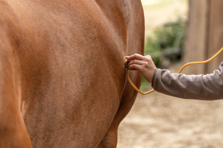
Evaluating movements with ultrasound provides functional data, increasing diagnostic accuracy. The equine body is in constant motion (even when we don’t want it to be), and this motion allows us to evaluate the moving parts, and the way they work together in the body.
Other than fluoroscopy, no other imaging modality can evaluate tissues in motion, and fluoroscopy is limited to evaluating bony structures, not soft tissue. Ultrasound can evaluate accessible soft tissue structures, and it is also the most sensitive tool for evaluating bone surface detail. Magnetic resonance imaging (MRI) and computed tomography (CT), actually employ computer software specialists to write “motion correction” programs that eliminate its effect on imaging. If a horse moves when performing standard radiographs, we must repeat these views.
All ultrasound is performed in “real time” and most ultrasound devices allow acquisition of “dynamic” video clips. This allows assessment of any accessible anatomical structure that moves. This includes most joints, both large and small, both axial and appendicular. It is valuable to image joints with any range of motion—minimal such as with cervical articular process joints, and those with a large range of motion like the carpus and tarsus.
Dynamic ultrasound is also used to evaluate muscles and tendons, which functionally work together as a musculotendinous “unit,” but they can be evaluated together or separately. Ligaments do not create motion like the musculotendinous unit, but just like bones, they should be evaluated as they are moved.
Ligaments and tendons both have attachments onto bone surfaces (entheses) and these entheses can be observed as a ligament or tendon performs its action at the enthesis.
Dynamic sonography allows fine-tuned assessment of soft tissue relationships. The distal palmar/plantar metacarpal/metatarsal soft tissues (superficial digital flexor tendon (SDFT), deep digital flexor tendon (DDFT), palmar/plantar annular ligament, palmar/plantar (intersesamoidean) ligament, palmar/plantar metacarpal/metatarsal fascia, digital tendon sheath and the mannica flexorum) are the best examples. (The mannica flexorum is a band-like structure of the superficial digital flexor tending that wraps around the deep digital flexor tending just above a horse’s hock.)
Subtle restrictive adhesions that reduce the normal “gliding” mechanism between these structures can be visualized. Fibrillation, synovial proliferation and small tears are more accurately identified. Fiber and fibrocartilage tearing of tendons and ligaments occurs when forces override their normal stress/strain capacity. This tearing might have directionality.
Most tendon and ligament injuries can be observed in transectional (cross section) imaging; however, they can also sustain longitudinal damage, and this can be difficult to capture sonographically.
Dynamic imaging—specifically flexing and extending ligaments and tendons—can allow a “spreading” of these longitudinal tears, allowing us to define these tears. These are most commonly observed in the deep digital flexor tendon, suspensory branches and intermediate patellar ligament.
Take-Home Message
Becoming familiar with dynamic functional sonography techniques is an invaluable tool for the expanding musculoskeletal ultrasound toolchest. Dynamic imaging should be practiced on normal structures on a constant basis to allow a practitioner to quickly develop an awareness of what is normal and abnormal. The more we use these tools, the more they become “second nature.” We also develop a more keen awareness of functional anatomy.
About the Author
Cooper Williams, VMD, DACVSMR, Certified ISELP Instructor, grew up in Cecil County Maryland, where he learned to play polo and train polo ponies with his family. He ultimately obtained a five goal rating and played professionally for five years. In the summer of 1981, he was a member of the United States polo team that competed throughout England. He coached the Valley Forge Military Academy polo team from 1981 to 1985. In 1984, he graduated with honors from the University of Pennsylvania School of Veterinary Medicine. He accepted a post graduate internship at Delaware Equine Center, where he developed their ultrasound imaging program. In 1987, he moved to Maryland where he worked for two years at Maryland Equine Center, also developing their ultrasound program. In 1989, Williams started his own practice, focusing on diagnostics and sports medicine in equine athletes. After three years of rigorous study, learning in depth about advanced diagnostic imaging, Williams passed his examination and was certified by the International Society of Equine Locomotor Pathology (ISELP) in 2009 and has continued on as a certified instructor with ISELP, teaching other equine veterinarians all over the world. Cooper is one of a small group of veterinarians in the world who is certified by the International Society of Equine Locomotor Pathology in advanced ultrasound imaging. In August 2014, Williams successfully passed his examination and is officially a Diplomate of the American College of Veterinary Sports Medicine and Rehabilitation. There are only a small group of veterinarians in the world that now can claim they are a specialist in sports medicine and rehabilitation. He is an active member of the North American Regenerative Medicine Association as well as being on the veterinary advisory board for ACell, Inc., a tissue engineering company.
Williams is a paid advisor for Butterfly Network.
![[Aggregator] Downloaded image for imported item #19769](https://s3.amazonaws.com/wp-s3-equimanagement.com/wp-content/uploads/2026/05/31141823/EDCC-Unbranded-11-scaled-1-768x512.jpeg)

