Equine practitioners commonly encounter patients with masses, nodules, and swellings in varying sizes and locations. During a Burst session at the 2025 American Association of Equine Practitioners Convention, Lindsay Deacon, DVM, of Littleton Equine Medical Center, shared her steps for assessing them.
Ultrasound and Documentation
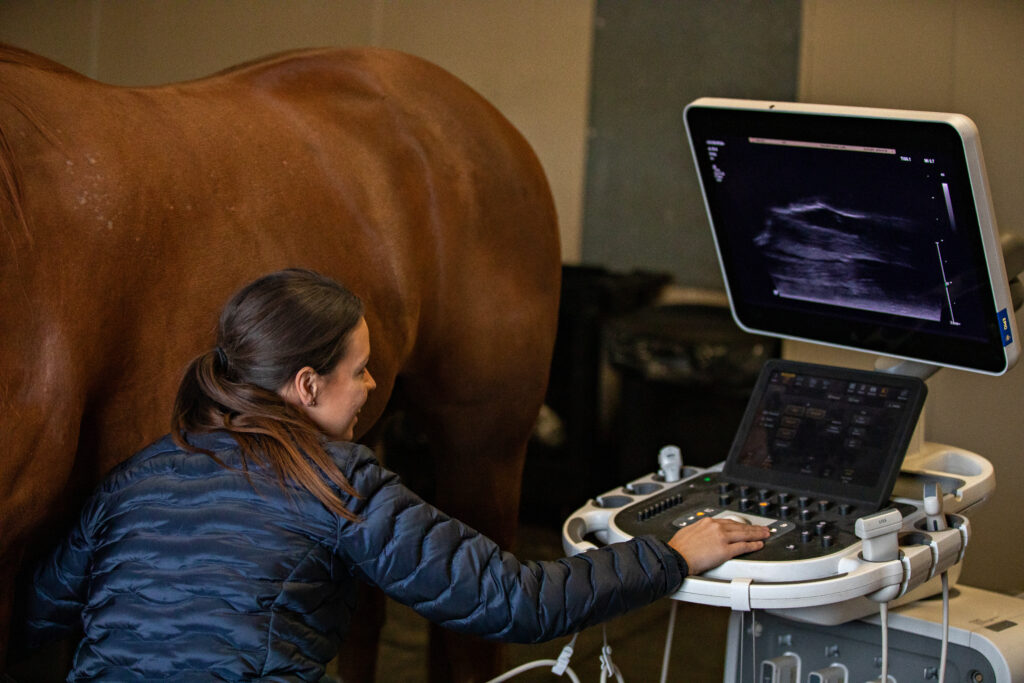
The first diagnostic of choice in most of these cases is ultrasound. Deacon recommended using both a curvilinear and a linear transducer.
“A lower frequency (2-5 MHz) curvilinear transducer can be used as a starting point or to get an overall big picture of the extent of pathology,” she explained. “A higher frequency (7-18 MHz) linear transducer is then used to assess the area in more detail.”
She emphasized the importance of knowing normal anatomy and documenting the normal regional structures on ultrasound. “It’s helpful to label these images on your ultrasound, especially complicated images, so someone else rechecking the horse would be able to understand what you’re looking at,” she said.
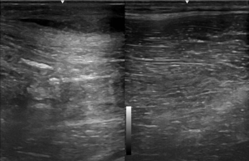
When in doubt, said Deacon, remember the horse has two sides: “An abnormality might be more apparent when comparing it to the normal opposite side.”
Determine What You Don’t Know
Once you’ve mapped out the normal structures on ultrasound, move on to what you don’t know, she said. Characterize the mass ultrasonographically using size (measurements of length, width, and depth of the mass to check progress over time), shape, margins, and echogenicity.
To determine the shape of the mass, rotate your ultrasound transducer on the structure of interest (e.g., orthogonal views). “This can help confirm if the mass is spherical versus tubular in shape.”
When assessing the margins of a mass or swelling, pay attention to whether or not it has a capsule, she added, which is indicative of an abscess.
“The character of the mass or swelling contents is a critical piece of information that will help you narrow down your list of differentials,” said Deacon. “Pay attention to whether it’s homogenous vs. heterogenous, anechoic/hypoechoic/hyperechoic, solid vs. fluid. Color flow doppler can be used to assess for blood flow within the structure, which is particularly important for things like neoplastic masses or a vessel bleeding into a hematoma.”
Other Diagnostics
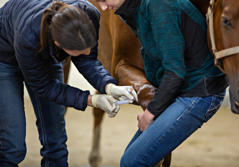
Characterizing the mass or swelling will help you narrow down a list of differential diagnoses so you can determine which additional diagnostics (e.g., radiographs, biopsy/aspirate, advanced imaging) you need to perform. It can also help you gather pertinent information that will influence the horse’s treatment and prognosis. “The steps prior to this point will also help determine your approach for ultrasound-guided drainage or aspiration, if indicated,” Deacon added.
Monitoring
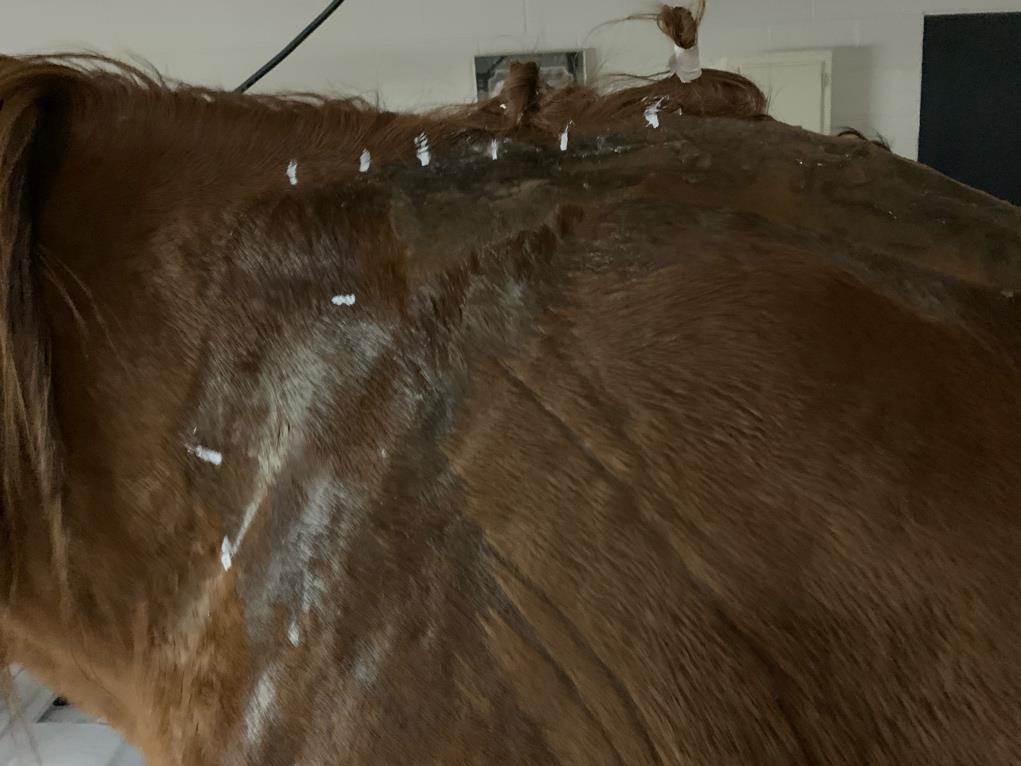
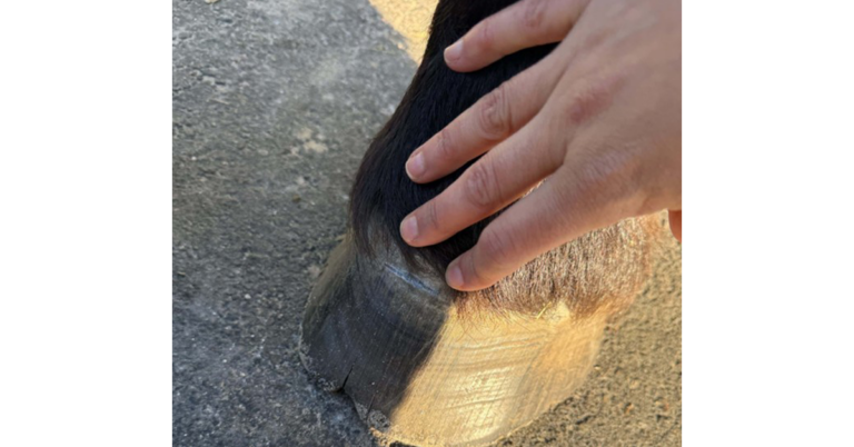
The final step in many of these cases is serial monitoring. “Be creative with this,” she said. “Utilize things like clipping, White-Out, permanent markers, rulers, or string to measure the initial size of a mass or swelling and help your owners monitor it over time,” in addition to ultrasound measurements.
Related Reading
- Rehabilitating Equine Soft Tissue Injuries
- Ultrasounding the Horse’s Medial Cranial Meniscotibial Ligament
- Optimizing Equine Ultrasound Machine Settings for Use in the Field
Stay in the know! Sign up for EquiManagement’s FREE weekly newsletters to get the latest equine research, disease alerts, and vet practice updates delivered straight to your inbox.
![[Aggregator] Downloaded image for imported item #18374](https://s3.amazonaws.com/wp-s3-equimanagement.com/wp-content/uploads/2025/09/30140012/EDCC-Unbranded-28-scaled-1-768x512.jpeg)