Ins and Outs of Equine Dental Extractions

The indications, techniques, complications, and referral recommendations for cheek teeth extractions.

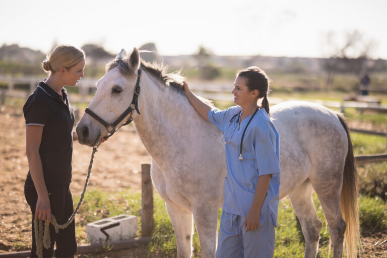
This article originally appeared in the Spring 2026 issue of EquiManagement. Sign up here for a FREE subscription to EquiManagement’s quarterly digital or print magazine and any special issues.

Veterinarian examining a horse's teeth prior to extraction.
Cheek teeth can present with various features that complicate extraction. | Shelley Paulson

Dental disease occurs with relative frequency in horses, necessitating that even gift horses require routine oral examinations. Cheek teeth pathology, such as periapical abscesses, tooth fractures, nonvital teeth, malocclusions, supernumerary teeth, or tooth resorption, might dictate extractions. And while these procedures can seem straightforward, many don’t go quite as planned.

Amelie McAndrews, DVM, DAVDC-EQ, owner of Garden State Equine Dentistry in Princeton, New Jersey, and clinical associate at the University of Pennsylvania’s New Bolton Center, says cheek teeth can present with the following features that complicate extraction:

  • Having curved (dilacerated) roots that anchor the tooth into the socket.
  • Presence of fractured crown, a tooth that is worn due to geriatric attrition and has minimal clinical crown, or malocclusions, making it difficult to grasp the tooth.
  • Pre-extraction radiographic evidence showing crown lucency, indicating the tooth is going to fracture.
  • Indications for catastrophic mandibular fracture based on radiographs.
  • Radiographic evidence of external replacement resorption, indicating the tooth will be cemented to the surrounding bone and very difficult to remove, likely accompanied by crown fracture.
  • Mesial or distal drift of surrounding teeth, causing crown interlock.

Alexis Leps, DVM, DEVDC-EQ, owner of LepsVetDent in France, adds that despite preoperative planning, “correctly predicting if simple oral extractions will be successful remains difficult.”

Preoperative Imaging Guides Extractions

One way to improve success is with proper surgical planning.

Leps says perioperative planning with an endoscopic oral examination, X-rays, and, when required, computed tomography (CT) are prerequisites that will guide the oral surgeon when deciding how best to extract a tooth.

“Dental X-rays are mandatory prior to performing any extraction,” she says. “The clinical crown (visible in the mouth) only represents between 10% and 30% of the tooth depending on the age of the patient.”

McAndrews concurs: “Radiographs are needed to make a diagnosis and for surgical planning, making sure none of the conditions noted above are present that will make tooth extraction more difficult.”

Diagnostic imaging prior to extraction can also elucidate whether extraction is indicated in the first place. “Just because a tooth looks ‘odd’ doesn’t mean it always needs extracting,” says Jessica Hunt, DVM, DAVDC (Equine), from Midwest Veterinary Dental Services, in Wisconsin.

“When radiographs do not provide adequate information about how a particular tooth is looking in three dimensions or in cases where sinusitis impairs radiographic interpretation of dental apices, CT is indicated,” says Leps, citing a study by Manso-Díaz et al. (2015).

Intraoperative Adaptability

Even with preoperative planning, practitioners must be able to quickly adapt intraoperatively.

“With experience, practitioners will be able to quickly determine that something with plan A, a standard oral extraction technique, is not working,” says Leps. “In such cases, practitioners need to move to plan B, C, and D, choosing the best plan according to the situation.”

She and colleagues from Ghent University in Belgium demonstrated this extraction agility as part of a study. They reviewed medical records from horses presenting for equine cheek extractions between October 2020 and July 2023. In total, 461 cheek teeth were extracted from 376 horses, including 292 maxillary and 169 mandibular cheek teeth.

A standard extraction technique was employed in cases with sufficient clinical crown and was successful in 77.2% of cases.

“This shows that even ‘standard extraction techniques’ aren’t simple, routine, or standard,” Leps explains. “Dental extractions are challenging surgeries that require good surgical planning, good anatomical knowledge, and a wide range of dedicated tools. Anything can go wrong and, as you can see, 22.8% of extractions in our study required a more advanced technique to successfully remove them.”

In the 22.8% of extractions that were not amenable to “standard” extractions techniques, an intraoperative decision was made to move to a more advanced extraction technique. In that study, Leps et al. used sectioning in 29 of the 461 extractions (6.3%). Most of those teeth were maxillary teeth sectioned into two or three pieces.

Reasons leading to sectioning included:

  • Very decayed teeth with pulp exposure, multiple infundibular dental caries, and complicated crown fracture.
  • Crumbling of the crown when molar spreaders were applied.
  • Unsuccessful application of minimally invasive transbuccal screw extraction (MITSE) technique.

The sectioning technique involved first lavaging the buccal cavity thoroughly. Using 3.175 millimeters double cut carbide burrs of varying lengths (38-77 millimeters), maxillary teeth palatal roots were separated from the buccal roots and burring was performed until bleeding periodontal ligament became visible. If the tooth sections still could not be removed, then a second cut was made to separate the two buccal roots. The alveolus was checked after removal to ensure no remaining tooth fragments persisted. For mandibular cheek teeth, sectioning was performed halfway along the mesiodistal length of the tooth. Radiographic guidance was used to ensure proper trajectory of the cut. Once cut appropriately, periodontal ligament luxation was performed and the two fragments individually elevated.

Mean surgical time in those 29 extractions was 106.6 minutes (range 30-235 minutes). The extraction was sufficiently challenging in three horses that the procedure needed to be completed under general anesthesia.

While complications occurred in one-quarter of the cases, “overall, complications associated with dental sectioning are low,” says Leps.

This is particularly true compared to repulsion of cheek teeth through sinuses, which carries a complication rate of 40-80%.

“Retropulsion has a high complication rate and should not be performed by general practitioners,” says McAndrews. “I think an important skill for general practitioners is to get a sense for which teeth are likely to be successful oral extractions and which teeth might need additional procedures to extracted and when to refer.”

Delving Into Complications

In 2022, Padraic Dixon, MVB, PhD, DECVD(Eq), FRCVS, a private surgical consultant from Edinburgh, published a report discussing complications associated with cheek teeth extractions, noting they occur even in the hands of the most skilled veterinarians. Some complications he highlighted included:

  • Damage to adjacent teeth causing pulp exposure and subsequent apical infection. Further, if the blood supply to the apical aspect of the tooth is inadvertently damaged, the tooth might become devitalized and later infected or fractured.
  • Traumatic alveolar damage and supporting bones leading to sequestration, osteomyelitis, and local soft-tissue infection.
  • Retained dental fragments that remain attached to the alveolar wall. Post-extraction/follow-up dental radiographs can identify these fragments, allowing practitioners to remove them.
  • Infraorbital nerve injury, resulting in violent headshaking for about one to two weeks post-extraction. Typically occurring in younger horses, this condition resolves with acepromazine therapy rather than corticosteroids or non-steroidal anti-inflammatories.
  • Surgical site infections at trephine repulsion sites promoting nonhealing tracts, which require practitioners to look for intra-alveolar dental or bone fragments. Infections of the alveolar bone or osteomyelitis of the supporting bones can also occur. This is particularly true when apical infections were present and the main indication for extraction.
  • Oromaxillary or oronasal fistulae, recognized by food-containing nasal discharge post-extraction, can develop following damage to the alveolar apex.
  • “Dry socket” or nonhealing alveolus that remains attached to the alveolar bone occurs because of inadequate formation of a blood clot that normally protects the bone. Time and packing of exposed alveolar bone will allow the underlying healthy bone to become covered in granulation tissue to permit healing.

“In my opinion, retropulsion should be avoided at all costs in the field, remaining in the hands of specialists,” says Leps.

Surgical Planning, Patient Preparation, and Cooperation

Patient preparation is equally important as the extraction itself.

In Leps’ study, for example, all horses underwent oral endoscopy prior to the procedure, together with standard dental radiographs (left and right dorsal to ventral 30-degree oblique projections for maxillary cheek teeth and left and right central to dorsal 45-degree oblique projections for mandibular cheek teeth).

Horses in Leps’ study were sedated with acepromazine, detomidine, and morphine intravenously and maintained with a continuous rate infusion of detomidine. Midazolam was administered as needed to minimize masticatory movements.

“Appropriately using neuroleptanalgesia and regional anesthesia improves the success rate in dental extractions,” she says.

“Regional nerve blocks are the key to success when performing dental surgery in the standing sedated horse,” Hunt adds. In her hands, heavily sedated patients benefit from maxillary and infraorbital nerve blocks as well as mandibular and inferior alveolar nerve blocks.

“Patients need to be sedated to perform these blocks to prevent sudden head movements that can traumatize the neurovasculature and the injection site antiseptically prepared to reduce the chances of infection,” says Hunt. “Patients can become very reactive even while heavily sedated. The infraorbital and mental foramen nerve blocks tend to result in the most reactivity, due to the proximity of the needle to the nerve bundles.”

She recommends selecting local anesthesia for blocks based on time of onset and duration of action. Lidocaine has a rapid time of onset (one to two minutes) but only lasts for one to two hours. Bupivacaine, however, lasts three to eight horse, with an onset of action of five to 10 minutes.

“Select the smallest needle gauge possible that will penetrate the skin but not traumatize the neurovascular structures,” advises Hunt. “I like Tuohy needles that have rounded bevels with the cutting edges offset to the side for maxillary nerve blocks. I use a 25-27-gauge, 1.5-inch needles for infraorbital nerve blocks.”

During her presentation at the 2024 Annual Convention of the American Association of Equine Practitioners, Hunt gave detailed instructions for performing various blocks. The abstract describes the following relevant blocks for removing cheek teeth:

Maxillary nerve block to desensitize all the ipsilateral maxillary cheek teeth, gingiva, and facial soft tissues rostral to the infraorbital foramen.

“The lacrimal, major and minor palatine, and caudal nasal nerves may inadvertently become anesthetized as well necessitating eye lubrication during the procedure,” she says.

To perform this block, follow the facial crest caudally and extend 2-3 centimeters (up to 1.5 inches) past the dorsal reflection of the zygomatic arch. Insert the needle perpendicular to the skin, advancing it until you feel a “pop,” then an additional 5 millimeters (0.2 inches). Hunt recommends injecting 10 milliliters of local anesthetic at this location.

Infraorbital neve block to desensitize the ipsilateral maxillary premolars, gingiva, and facial soft tissue (lips, nose, and skin). If the needle advances sufficiently far, the molars might also be blocked.

“In my surgical experience, you can desensitize all maxillary cheek teeth with this block, but there is no paper supporting this as far as I am aware,” says Hunt.

To perform this block, palpate the nasoincisive notch and rostral edge of the facial crest while dorsally displacing the levator nasolabialis muscle. The infraorbital foramen can be palpated here. Inject 3-5 milliliters of local anesthetic.

Inferior alveolar block to desensitize the mandibular nerve and the ipsilateral mandibular alveolar mucosa, teeth, gingiva, skin, and labial mucosa. This nerve can be approached intra- or extraorally. From the extraoral approach, hold a 20-gauge, 6 Tuohy needle, or 12.5-centimeter spinal needle against the skin and insert it 1-2 centimeters medial to the ventromedial aspect of the mandible.

“The external landmarks are the ‘imaginary lines’ made along the occlusal surface of the mandibular cheek teeth and a second line from the lateral canthus of the eye ventrally to the mandible,” Hunt explains. “The intersection of these two lines is used to estimate the location of the foramen, which lies on the medial aspect of the mandibular ramus. Inject 10 milliliters local anesthetic at this location.”

For the intraoral approach, the patient must be adequately sedated to prevent the tongue from moving. Using a 23-gauge, 19-25-millimeter butterfly catheter grasped with alligator forceps, introduce the needle into the oral mucosa overlying to rostral ramus just caudal to the mandibular third molar at the level of the occlusal surface. Inject 3-5 milliliters of local anesthetic.

Complications can occur following routine dental nerve blocks, from mild concerns such as local hematoma formation, regional infection, and failure to desensitize to more serious issues such as septic meningitis, neuropraxia, and facial trauma (e.g., secondary to inadvertently blocking nearby nerves such as the lacrimal or lingual nerves).

“Nonetheless, regional nerve blocks improve patient compliance, decrease procedure length, and provide a safe work environment,” says Hunt.

She further recommends repeating the nerve block at the end of the procedure for postoperative pain control.

Concluding Recommendations and Referrals

“It is always OK to refer patients prior to pursuing surgery,” says McAndrews. “Some oral extraction attempts will be unsuccessful, even for the most experienced of clinicians. If the practitioner doesn’t have the time, desire, skill, or instrumentation to perform the extraction, it is best to refer before attempting extraction.”

Leps agrees: “You don’t need to be in the position to abort intraoperatively. Overall, I believe it is better to put these kinds of surgeries in the hands of veterinarians who are trained to be able to successfully extract the tooth while being as minimally invasive as possible and always thinking about animal welfare first.”

“Referral is never wrong and there is potentially less trauma to the patient and fewer complications when referral is pursued in difficult cases,” says McAndrews.

Stay in the know! Sign up for EquiManagement’s FREE weekly newsletters to get the latest equine research, disease alerts, and vet practice updates delivered straight to your inbox.

categories
tags
Trending Articles
[Aggregator] Downloaded image for imported item #19998
Strangles Case Confirmed in King County, Washington
Female Vet Giving Injection To Horse In Stable
Early Career Insights: How to Build Trust With Horse Owners
[Aggregator] Downloaded image for imported item #19998
Strangles Cases Confirmed in 2 Florida Counties
Equine Veterinarian Exam Using an Ultrasound
Business Briefs: What Is Your Practice's Competitive Advantage?
POLLS AND SURVEYS
Newsletter
Get the best from EquiManagement delivered straight to your inbox once a week! Topics include horse care, disease alerts, and vet practitioner updates.

"*" indicates required fields

This field is for validation purposes and should be left unchanged.
Name*
Country*

Additional Offers

Untitled