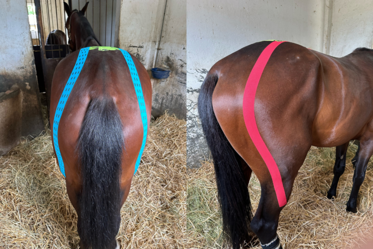
Veterinarians have made huge strides in diagnosing and treating pathology involving the caudal cervical region, the junction of the seventh cervical vertebra and the first thoracic vertebra (C7-T1)—once considered untreatable.
According to John Janicek, DVM, MS, Dipl. ACVS. of Brazos Valley Equine Hospitals in Salado, Texas, pathology at C7-T1, most commonly but not always caused by cervical vertebral stenotic myelopathy (CVSM), has gained tremendous traction.
Clinical Signs of Caudal Cervical (C7-T1) Pathology in Horses
Caudal cervical pathology is now recognized as a common cause of pain, lameness, ataxia, and poor performance. Affected horses have uni- or bilateral atrophy of the pectoral muscles, including the brachiocephalicus and omotransversarius as well as the antebrachial muscle, Janicek explained during his presentation at the 2023 American Association of Equine Practitioners Convention, held Nov. 29 to Dec. 6 in San Diego, California.
Further, horses with caudal cervical pathology such as CVSM might:
- Have difficulty eating off the ground.
- Struggle to walk in circles.
- Resist moving forward with the head and neck elevated.
- Drag all four toes when backing up, especially the forelimbs.
- Have amplified gait abnormalities when ridden.
Diagnostic Imaging for C7-T1 Lesions in Horses
“Lesions of the caudal cervical spine, C7-T1, can often be missed on routine radiographs,” Janicek said. “It’s important in these cases to perform a caudal retraction of the limb that is ipsilateral to the generator. This eliminates superimposition of the scapula, scapulohumeral, and shoulder musculature, removing it from the primary view.”
While dorsal spinal cord compression at C7-T1 can potentially be seen on standard myelography, CT comes in handy and has big benefits because it allows for imaging in the coronal, sagittal, and axial axis. In fact, said Janicek, CT myelography should be considered the gold standard for diagnostic imaging in cases of caudal cervical pain, which can now be performed under general anesthesia as far caudally as T4/5.
Ventral Interbody Fusion
Janicek said intervertebral foramen stenosis has also gained a lot of traction, as has disc degeneration, which never used to be mentioned as a problem. Veterinarians can treat all three of these conditions with ventral interbody fusion of the C7-T1 articulation, a procedure no longer considered a salvage operation.
However, Janicek did describe a few surgical obstacles when performing a ventral interbody fusion C7-T1.
“There are few anatomic identification markers of C7,” he explained. “There are no ventral spines palpable, only two bony tubercles. Careful dissection in that area is required because the carotid artery sits right there. Also, the sternum is large, making things awkward to work around at times.”
Demonstrating the value of ventral interbody fusion of C7-T1, Janicek presented data from 29 cases that underwent this procedure. Surgery was considered successful (e.g., the horses were used for riding or pasture turnout) in 22 of those cases (76%). Three horses were euthanized, two died, and two were lost to follow-up.
The convention proceedings included specific surgical tips, including suggestions regarding general anesthesia, head elevation, leaving the endotracheal tube in for a longer period than usual, and rope recovery systems.
Postoperative Rehabilitation
Postoperative rehabilitation must be tailored to each patient and depends on the level of ataxia before and after surgery. Dynamic mobilization can generally begin four months after surgery and includes lateral and ventral bending of the cervical spine to improve range of motion.
“Rehabilitation lets the horse regain their confidence in themselves and improves proprioception,” said Janicek. “Balance pads may facilitate this process.”
Final Thoughts
In sum, Janicek said ventral interbody fusion of C7-T1 is feasible for eliminating static and dynamic spinal cord compression and pain. The prognosis for recovery is good.
“The surgical goal is to return horses to athleticism and improve performance, but the intent is to improve comfort and quality of life,” said Janicek.
Related Reading
- Horse Back Pain Rehabilitation
- Managing Lumbosacroiliac Joint Region Pain in Horses
- Is That Horse’s Radiographic Neck Lesion Important?
Stay in the know! Sign up for EquiManagement’s FREE weekly newsletters to get the latest equine research, disease alerts, and vet practice updates delivered straight to your inbox.

![[Aggregator] Downloaded image for imported item #18375](https://s3.amazonaws.com/wp-s3-equimanagement.com/wp-content/uploads/2025/09/30140031/EDCC-Unbranded-26-scaled-1-768x512.jpeg)


