This article originally appeared in the Spring 2026 issue of EquiManagement. Sign up here for a FREE subscription to EquiManagement’s quarterly digital or print magazine and any special issues.
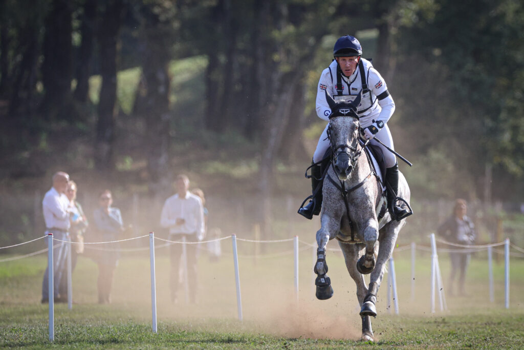
Emmanuelle van Erck-Westergren, DVM, PhD, ECEIM, ECVSMR, of Equine Sports Medicine Practice, in Belgium, described a comprehensive approach to working up non-orthopedic poor performance issues in English sport horses at the 2025 American Association of Equine Practitioners Convention.
Respiratory Issues
Sport horses spend significant time training, competing, and traveling—activities that increase the risk of inhaling dust and debris deep into the airways. Chronic stress related to campaigning and exposure to pathogens when co-mingling at events can further compromise immune function. Van Erck-Westergren explained that with training, muscles develop and the heart enlarges, but airway development usually lags behind. With this in mind, the first limiting factor to consider for non-orthopedic problems with equine performance is the respiratory system.
A horse’s oxygen transport capacity increases 50-fold between rest and exertion from 4 liters/minute to 200 liters/minute. The spleen aids in oxygen transport by releasing 30% of red blood cells into circulation. All the blood must circulate through the lungs, resulting in massive pressure on a horse’s lungs. The bigger the heart, the greater the pressure, she noted, increasing the risk of exercise-induced pulmonary hemorrhage (EIPH) with exertion.
In general, horses experience oxygen deficiencies due to narrow upper airways, compulsory nasal breathing, and rapid capillary transit time. As red blood cells pass through the lungs during exertion, they spend only a quarter of a second uploading oxygen and cannot entirely achieve a full load. In addition, when horses breathe during locomotion, they can only inhale when the limbs are in extension, followed by a diaphragmatic push for exhalation. As such, an exercising horse cannot exceed a certain rhythm, said van Erck-Westergren.
A Comprehensive and Discipline-Specific Work-Up
Van Erck-Westergren stressed that veterinarians understand the demands of specific disciplines when evaluating horses with poor performance. A trot-up does not necessarily indicate a horse is fit to perform, she said. It is better to gather blood samples for information, especially after exercise. A horse might experience a decrease in maximal oxygen consumption if it is anemic or if the heart beats irregularly. Bloodwork allows vets to check for anemia, white blood cell counts, leucopenia, acute phase proteins indicative of inflammation, and globulins. Muscle enzyme elevations suggest tying-up, lower airway disease, or cardiac arrhythmia.
It’s helpful to check vitamin E, magnesium, and selenium levels since they are essential for good muscle function. Gastrointestinal issues might affect calcium levels. Hepatic parameters provide information about feed safety; mycotoxins elevate liver enzymes, for example. Renal function evaluation is important if a horse receives bisphosphonates.
Van Erck-Westergren advised doing a comprehensive clinical exam for cardiac and respiratory function; fitness, stamina, and heart rate recovery; and locomotion. Watch under saddle for horse and rider interactions that could affect performance.
Exercise Testing
Field exercise testing allows vets to look at the whole horse performing its skill set. Behavior ethograms help elucidate ridden pain. A horse asked to hyperflex its neck might experience upper airway instability. In one study, researchers examined head position and airway and nasopharynx diameters and found that when a horse’s head is in a dorsal extended position, airway diameter increases by 66%.
Van Erck-Westergren said a change in bit type can also help improve airway openings and horse comfort. The larger and stiffer the bit, the more difficult it is for a horse’s tongue to find space. The tongue shifts upward and pushes the larynx back so it can no longer stay ventral and rostral in the oropharynx. Instead, it tilts backward to loosen the hyoid apparatus and make airway walls more flaccid, which decreases the airway opening.
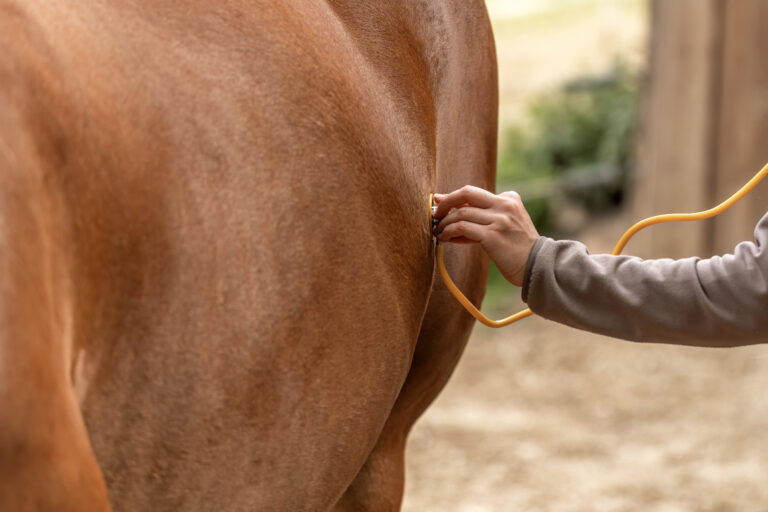
Another good testing method of the respiratory tract is endoscopy when horses are on a treadmill or a track. Measuring VO2 at exercise, lactate assessments, motion analysis, airway endoscopy, and an ECG on the heart are useful tools to identify a cause for poor performance.
Exercise testing allows veterinarians to measure heart rate and heart rate recovery. With aerobic exercise, pyruvic acid from blood glucose and glycogen breakdown enters the Krebs cycle in the mitochondria to produce ATP energy that drives muscle contractions. Because fatty acids increase the ATP produced, adding fat to the diet might be helpful. Yet with anaerobic metabolism, pyruvic acid transforms into lactic acid, which adversely impacts muscle contraction and results in fatigue. Lactic acid concentrations increase with cardiac problems or respiratory problems from lower airway disease, anemia, mitochondrial disease, or heat stress.
A noninvasive submaximal exercise test evaluates how well a horse uses oxygen for aerobic metabolism and helps detect subtle problems. These tests can also reveal whether a horse is experiencing lameness or other pain that increases the heart rate. A transtracheal wash and BAL provide information on cytology that could represent infection, inflammation, or EIPH. Cytology on tracheal samples is important for asthmatic horses so the vet knows if it’s safe to treat with corticosteroids.
Gastrointestinal Pain
Another potential cause of poor performance is gastrointestinal pain. When exercising with speed or exertion, a horse’s core abdominal muscles compress to alter pressure inside the stomach. In one study, researchers placed a pH probe to the stomach cardia and found that pH drops with exertional exercise as gastric acid at the bottom of the stomach splashes up onto the mucosa. There is an overall positive correlation between pain scores and equine glandular gastric disease (EGGD), equine squamous gastric disease (ESGD), and total equine gastric ulcer scores. A 12-week study assessing ridden pain ethogram scores for horses with and without gastric disease showed dietary changes rich in fiber and probiotics can alter gastric disease and improve behavior.
Related Reading
- Equine Rib Fractures as Cause of Poor Performance
- Is Gastric Disease Really to Blame for Equine Behavior Issues?
- Approach to Horses with Poor Performance
Stay in the know! Sign up for EquiManagement’s FREE weekly newsletters to get the latest equine research, disease alerts, and vet practice updates delivered straight to your inbox.
![[Aggregator] Downloaded image for imported item #19769](https://s3.amazonaws.com/wp-s3-equimanagement.com/wp-content/uploads/2026/05/31141823/EDCC-Unbranded-11-scaled-1-768x512.jpeg)

