“The medial cranial meniscotibial (MCMTL) is commonly implicated in cases with meniscal injury, so it’s very important to evaluate this ligament in all cases of stifle lameness,” advised Sherry Johnson, DVM, MS, Dipl. ACVSMR, senior partner at Equine Sports Medicine and Rehabilitation, LLC in Pilot Point, Texas/Scottsdale, Arizona, during a “Burst” session at the 2024 American Association of Equine Practitioners Convention.
The MCMTL originates from the cranial horn of the horse’s medial meniscus and inserts on the medial aspect of the tibial intercondylar eminence.
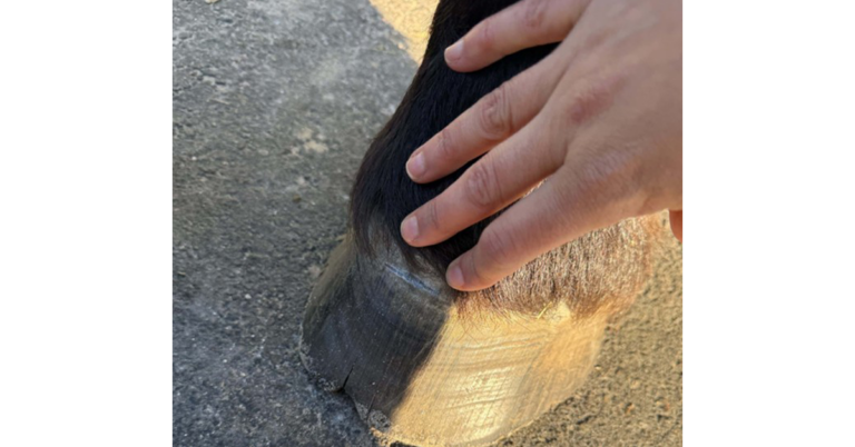
After blocking lameness to the stifle, wash and clip the region of interest before applying gel. Using a linear probe with optimized machine settings, face the stifle/tail and hold the limb in a non-weight-bearing position.
“Be careful not to overflex the joint, or it will close off the anatomic window you need to adequately visualize the MCMTL,” said Johnson.
First, locate the horse’s MCMTL in long axis but transverse to the medial meniscus. Center the image on the medial meniscus. Trace from the medial meniscus to the cranial horn and then toward its insertion onto the tibia. Hold the probe underhand in transverse, moving in an axial to abaxial direction. Note how the shape of the ligament changes along its length. It starts with a meniscus shape at the cranial horn. Then, the body becomes round. Finally, it becomes flat and thin at the tibial insertion.
“If you haven’t seen every shape, you haven’t conducted a thorough exam,” Johnson cautioned.
To get started in long axis, Johnson recommended placing the probe in the “sweet spot” between the medial and middle patellar ligaments with the limb held in non-weight-bearing.
“Start high and move the probe distally. Push in and rotate the probe counterclockwise just a little, and grip the probe in an overhand position for this portion of the exam,” she said.
Johnson offered two other pieces of advice for imaging the horse’s MCMTL:
- Small adjustments make a big difference in this study.
- Push hard on the probe.
“If your arm isn’t shaking by the end of the exam, you didn’t push hard enough!” she said.
Related Reading
- Rehabbing Equine Hind Proximal Suspensory Ligaments
- Optimizing Equine Ultrasound Machine Settings for Use in the Field
- Using Information from Cross-Sectional Imaging in Daily Practice
Stay in the know! Sign up for EquiManagement’s FREE weekly newsletters to get the latest equine research, disease alerts, and vet practice updates delivered straight to your inbox.
![[Aggregator] Downloaded image for imported item #18374](https://s3.amazonaws.com/wp-s3-equimanagement.com/wp-content/uploads/2025/09/30140012/EDCC-Unbranded-28-scaled-1-768x512.jpeg)