MRI has been the gold standard for diagnosing foot pain in horses. To minimize the chances of inconclusive examinations, however, veterinarians must select cases for MRI carefully.
During a lecture at the 2025 British Equine Veterinary Association Congress, Annamaria Nagy, DrMedVet, PhD, DACVSMR, DECVSMR, FRCVS, of the Equine Department and Clinic at the University of Veterinary Medicine Budapest, in Hungary, explained what to do if you don’t find abnormalities on an MRI for suspected foot pain.
“To clarify, negative MRI or MRI with no abnormalities means there are no abnormalities that explain the lameness and fit with the clinical picture,” she said. “In most horses, we can find something, and it can be tempting to assign minor lesions clinical significance.”
Nagy presented the three most common explanations for negative foot MRIs.
1. Imaging the Wrong Region
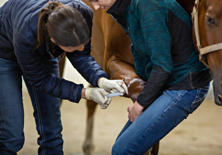
Ideally, the horse underwent a detailed clinical examination, gait assessment, and comprehensive diagnostic analgesia to identify the source of pain. Communication with the referring veterinarian is essential, including confirming the lame(r) limb, any previous diagnostic anesthesia, and when it was performed.
A chronically lame horse, for instance, that historically always blocked to the foot and has had repeated distal interphalangeal (coffin) joint medications, might not have had a nerve block in six months. A lot could have happened since then.
Also consider that some blocks, such as abaxial sesamoid and palmar digital nerve blocks, can alleviate or abolish fetlock and pastern region pain. “Therefore, a few screening sequences of the pastern region should be considered part of routine foot MRI examination,” said Nagy. “If images of the foot are inconclusive, the pastern and fetlock regions should be examined.”
2. MRI Is Not the Appropriate Imaging Modality
MRI might not be able to depict the lesion that’s causing pain, and computed tomography might be the more appropriate option. We know, for instance, that MRI does not illustrate hoof wall pathology well. MRI (especially low-field) can also miss small or mild osseous changes, such as fragments, mineralization, and resorptive subchondral bone injuries, said Nagy.
She recommended using CT to diagnose foot pain in cases where MRI did not identify significant abnormalities. “The combination of the two modalities can also guide the interpreter to mild MRI changes that might otherwise be missed or considered potential artefacts,” Nagy added.
3. Diagnostic Imaging Cannot Visualize the Source of Pain
In some cases, you simply won’t be able to visualize the source of pain with diagnostic imaging. These include poor foot conformation or trimming, pain in the heel region or digital cushion, mild solar bruising, and pain associated with equine metabolic syndrome.
“It’s important that we are prepared to go back to the basics, to our clinical examination if we need to after MRI, and make sure we haven’t missed anything,” Nagy said.
She added that MRI might also be negative if you’re too quick to pursue imaging—such as with very young horses, very recent onset of lameness, or subtle lameness.
In Summary
Faced with a negative MRI, “I think the most important take-home message is be prepared to rethink,” said Nagy. Critically reevaluate the horse’s history and the results of the clinical examination and repeat diagnostic analgesia if necessary.
She urged practitioners to resist the temptation to assign significance to minor lesions that do not fit with the clinical picture.
“If you’re absolutely sure the source of the pain is in the foot, we can consider further imaging, which will be CT in most cases,” she said.
Related Reading
- Daily Vet Life: Shifting Mindsets Surrounding Navicular
- Radiographs vs. MRI Show Limited Hoof Measurement Agreement
- Tips and Tricks to Acquiring Great Equine Foot Radiographs
Stay in the know! Sign up for EquiManagement’s FREE weekly newsletters to get the latest equine research, disease alerts, and vet practice updates delivered straight to your inbox.