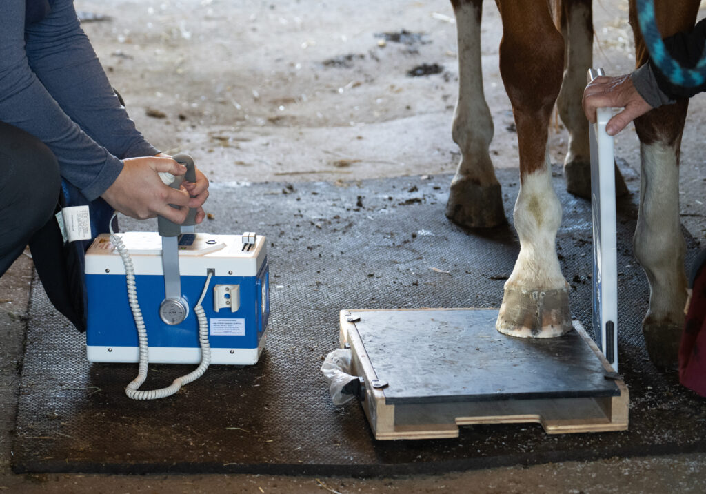
Veterinarians commonly use equine foot radiographs to measure anatomical conformation parameters. Comparison of measurements between radiographs and low-field magnetic resonance imaging (MRI) has not been extensively explored. A comparative cadaveric analytical study aimed to compare foot parameter measurements between radiographs and low-field MRI and to assess the effect of hoof wall markers on visualizing the hoof capsule (during MRI) and facilitating measurements.
Research Methodology
The researchers performed radiography and MRI on nine equine cadaver front feet with and without hoof wall markers (lead strips for radiography and a water-soaked hoof bandage for MRI). They calculated intraobserver reliability and intermodality agreement using intraclass correlation coefficients (ICC) with 95% confidence intervals (CI) and p < 0.05.
Research Findings
Intraobserver repeatability was generally good, apart from distal dermal frontal measurements. There was limited agreement between radiographic and MRI measurements. Results are presented as RAD indicating those obtained with radiography and T1, T2*, or STIR indicating those obtained with the relevant MRI sequence; m is added if a marker was used. Founder distance only showed good agreement for radiographic and T1 measurements with markers; ICC 0.78 (CI 0.33–0.95 p = 0.004). Intermodality comparisons for distal phalanx rotation were limited by intraobserver repeatability. Good agreement was noted for sole thickness and epidermal sole thickness measurements with markers; sole thickness (RADm vs. T1m ICC 0.81 [CI −0.04-0.96], p < 0.001; RADm vs. T2*m ICC 0.86 [CI 0.51–0.97], p < 0.001; RADm vs. STIRm ICC 0.91 [CI 0.66–0.98], p < 0.001) and epidermal sole thickness (RADm vs. T1m ICC 0.88 [CI 0.55–0.97], p < 0.001; RADm vs. T2*m ICC 0.83 [CI 0.41–0.96], p = 0.002; RADm vs. STIRm ICC 0.80 [CI 0.31–0.95], p = 0.004). Radiographic measurements with and without markers often had good to excellent agreement; for some parameters, hoof wall markers were associated with reduced intraobserver repeatability. The water-soaked hoof bandage aided MRI hoof capsule visualization; limitations included reduced repeatability and unattainable distal measurements.
Bottom Line
The limited agreement between radiographic and MRI measurements suggests these modalities are not interchangeable in equine foot assessment and that MRI hoof measurements cannot be used as a substitute for radiographic assessment. Hoof wall markers do not benefit foot measurements for either modality and might hinder radiographic interpretation.
https://beva.onlinelibrary.wiley.com/doi/10.1111/evj.14536
Related Reading
- Tips and Tricks to Acquiring Great Equine Foot Radiographs
- Disease Du Jour: Negative Palmar Angle in Horses
- The History and Future of MRI
Stay in the know! Sign up for EquiManagement’s FREE weekly newsletters to get the latest equine research, disease alerts, and vet practice updates delivered straight to your inbox.