This article originally appeared in the Winter 2024 issue of EquiManagement. Sign up here for a FREE subscription to EquiManagement’s quarterly digital or print magazine and any special issues.
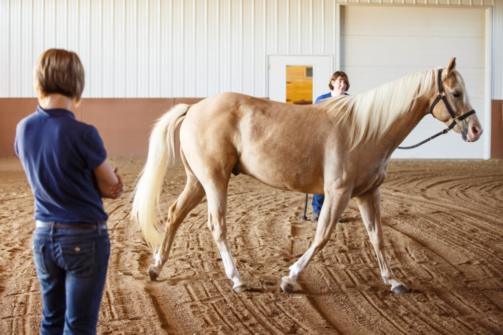
Forelimb lameness caused by pain isolated to the caudal heel negatively affects horses’ athletic performance and welfare. Historically, forelimb heel pain that blocked out using a palmar digital nerve block was quickly attributed to degeneration of the navicular bone, leading to a navicular syndrome diagnosis. We now know navicular syndrome affects more than just the bone and that caudal heel pain can stem from degeneration and inflammation of any structures comprising the podotrochlear apparatus. Hence, the term podotrochlosis has replaced the ambiguous navicular syndrome, giving a more encompassing view of the cause of lameness.
With the modernization of the name came advances in diagnosis as well as treatment. In this article, we’ll review the podotrochlear apparatus, the presentation of podotrochlosis, and relevant information from recently published articles, including the use of various imaging modalities and osteostixis for treating affected horses.
The Podotrochlear Apparatus and its Degeneration
The key players in the podotrochlear apparatus include the navicular bone, the collateral suspensory ligaments of the navicular bone, the distal sesamoidean impar ligament, the navicular bursa, and the deep digital flexor tendon.
“The navicular bone is a sesamoid bone with many important functions,” explains Anton E. Fürst, Dr med veta, DECVS, head of the division of equine surgery of the Vetsuisse Faculty, University of Zurich. “Firstly, the navicular bone, together with the podotrochlear bursa, acts as a sliding bearing (hypomochlion) for the deep flexor tendon. This allows the deep flexor tendon to move the coffin bone with less force (pulley principle). The navicular bone ensures that the deep flexor tendon attaches to the coffin bone at a constant angle in every position of the distal limb.”
He adds, “The navicular bone anatomically and functionally enlarges the articular surface of the hoof joint and, together with the hoof joint, plays an important role in weight-bearing in the various gaits of the horse. Finally, the navicular bone also has a shock-absorbing function. When the deep flexor tendon is loaded, the navicular bone is compressed.”
Podotrochlosis affects approximately 10% of the equine population, primarily Quarter Horses, Thoroughbreds, and warmbloods. Scientists suspect a genetic component in warmbloods, but so far the exact cause of this condition is largely unknown. Various theories exist to explain the pathophysiology of podotrochlosis. In Fürst’s 2021 article on foot problems co-authored with Christopher Lischer, Dr.Med.Vet., DECVS, Assoc.Member ECVDI, MRCVS, some of those theories include the following:
Vascular compromise. “Pathological anatomical examinations revealed thrombotically altered primary arteries, which led to compensatory vascularization of the navicular bone and subsequently to ischemic necrosis of the bone,” explains Fürst. “These ischemic changes are also considered to be the cause of the pain and lameness. However, many doubt this pathogenesis.”
Biomechanical abnormalities. “According to this theory, the degenerative processes on the navicular bone are caused by excessive compression forces on the distal part of the navicular bone, which is trapped in this area between the deep flexor tendon and the coffin bone,” says Fürst. “Excessive loading, malpositioning, and an unfavorable hoof shape lead over time to cartilage changes in the flexor facies and subsequently to changes in the subchondral bone and medulla of the navicular bone.”
Alternatively, a combination of these theories might explain the pathogenesis. In one of his publications, Fürst wrote, “Repetitive loading with nonphysiologic biomechanical forces combined with impaired venous drainage causes venous hypertension in the bone marrow resulting in intraosseous hypertension and bone edema. This compartment syndromelike condition triggers the accumulation of osmotically active proteins in the subchondral bone, which is characterized by increased tissue pressure, acidosis, pain, and a vicious circle of progressive pathologic changes.”
Foot conformation, as well as the activities the horse performs, might also contribute to podotrochlosis. These, says Fürst, might increase biomechanical forces in the podotrochlear apparatus.
Gold Standard Imaging Options for Podotrochlosis
Radiography remains the first-line diagnostic modality to assess navicular bone changes.
“When overt changes are present on radiographs, such as resorption of the bone, a diagnosis of navicular disease is established,” says Mathieu Spriet, DVM, MS, DACVR, DECVDI, DACVR-EDI, Professor of Diagnostic Imaging, School of Veterinary Medicine, Department of Surgical and Radiological Sciences, University of California, Davis. “If the radiographs remain within normal limits or are equivocal, then advanced imaging is indicated.”
For example, if enlarged synovial invaginations, enthesophytes, osteophytes, and/or sclerosis are present, additional imaging should be performed, preferably magnetic resonance imaging (MRI).
In fact, experts widely agree MRI is the gold standard for diagnosing podotrochlosis.
“MRI provides the most comprehensive assessment of both the navicular bone and associated ligaments and tendons,” says Spriet.
Emerging Diagnostic Options
Researchers are exploring other available diagnostic imaging techniques for podotrochlosis.
Computed Tomography
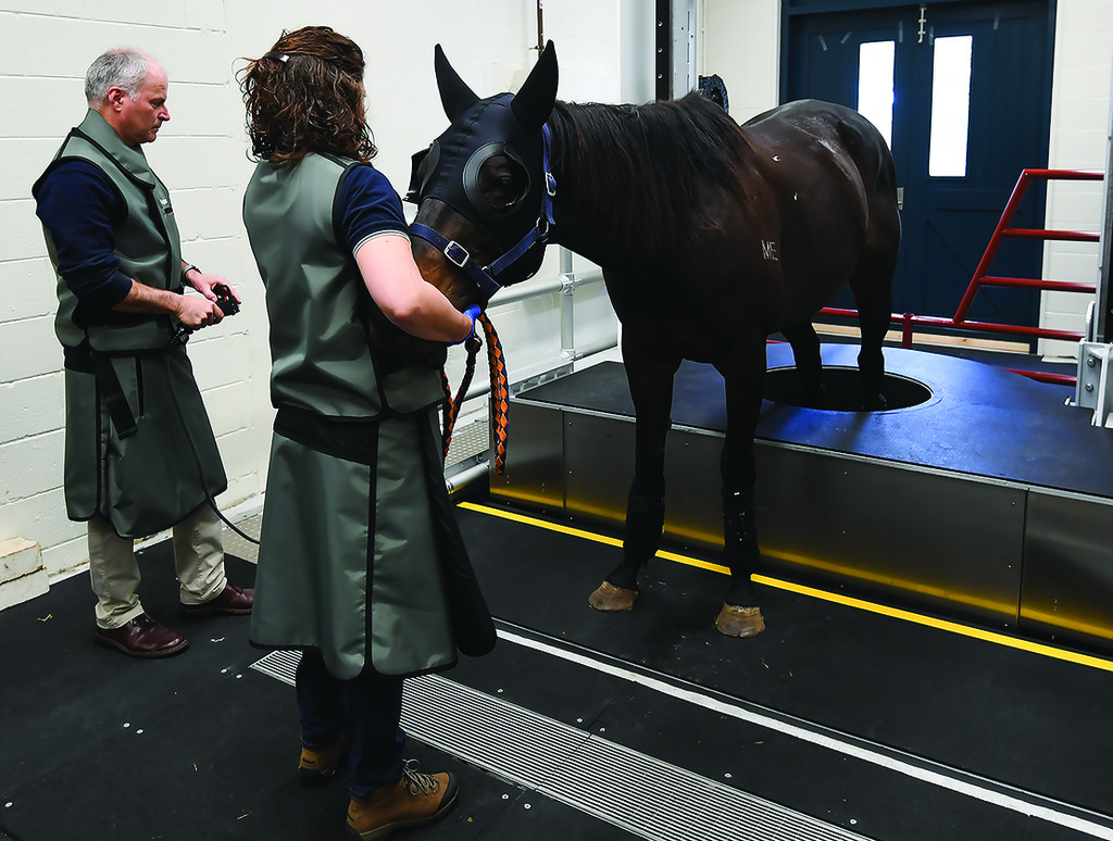
Sabrina Brounts, DVM, PhD, DACVS, DACVSMR, from the Department of Surgical Sciences, School of Veterinary Medicine at the University of Wisconsin, Madison, and colleagues recently studied a helical fan-beam computed tomography (CT) system’s ability to diagnose distal limb issues in standing, sedated horses (2022).
“There are two ways CT imaging can occur,” she explains. “One is cone-beam system CT imaging, and the other way is helical fan beam system CT imaging, with helical fan being more popular.”
Fan-beam CT’s benefits include:
- Excellent image quality with better soft tissue differentiation compared to cone beam.
- Less radiation dose for the patient than cone beam.
- No effect on image quality due to motion.
- Increased contrast resolution.
- Fewer artifacts.
In their study, Brounts et al. used the equine unit from AstoCT to scan the distal limbs of 167 standing, sedated lame horses in a two-year period. It has the ability to scan both limbs simultaneously in a natural weight-bearing position with short acquisition time.
“Scanning the limbs takes 15 seconds from fetlock down and 30-45 seconds from carpus/tarsus down. It is a very fast process,” she notes. “The longer procedure time is the time to get the horse ready, sedated, placed into the room with the scanner, scan, and walk the horse out again to the stall once all done. In most cases a procedure takes about 20 to 30 minutes (average 25 minutes), and we can scan three to four horses in an hour right now because we have perfected the routine.”
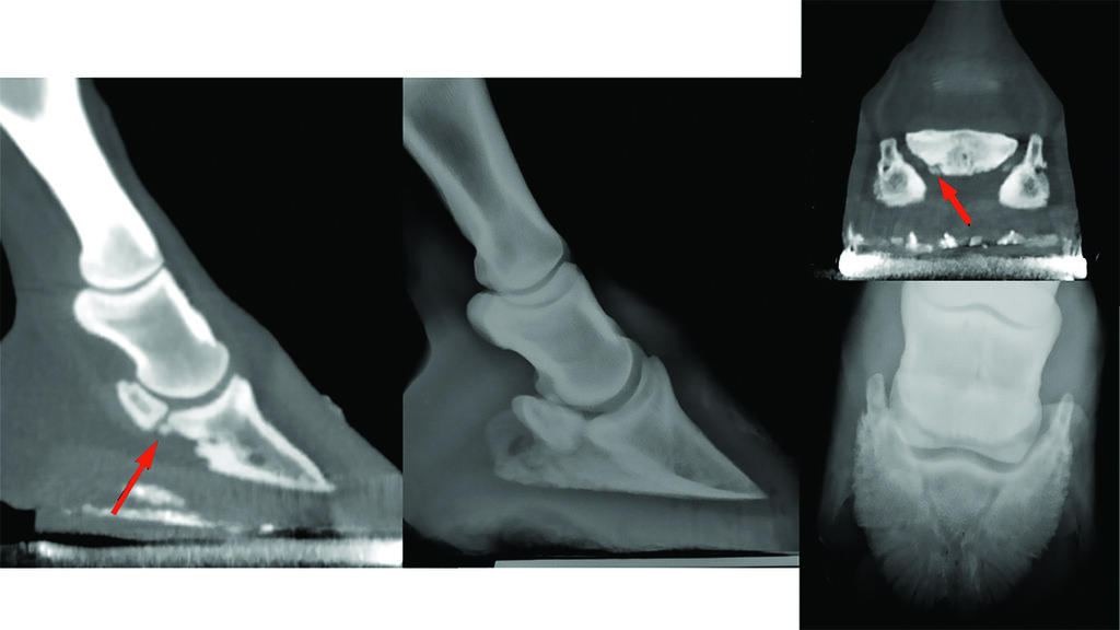
Brounts’ team obtained diagnostic scans from all study horses and localized lameness to the foot or pastern in 53% of cases. Almost half of those horses were diagnosed with navicular disease. Other causes of foot pain in the study included foot abscess, fracture of the distal phalanx, keratoma, osteoarthritis, pedal osteitis, fracture of the middle phalanx or navicular bone, and septic tendon sheath.
“If standing low-field MRI is used to image the podotrochlear apparatus, it should be accompanied by an additional imaging modality for a complete evaluation of anatomic structures,” they wrote.
“Low-field standing MRI has limitations such as detection of articular cartilage lesions in absence of subchondral pathology and imaging some anatomical structures in the pastern area,” Brounts explains. “So, if standing low-field MRI is used to image the podotrochlear apparatus, it would be wise to use an additional imaging modality.”
If you use high-field MRI for diagnosis, an additional modality is not necessary; however, high-field MRI can only be acquired under general anesthesia.
“Standing CT and PET/CT imaging might be the future for podotrochlosis horses, especially because these are imaging modalities that are becoming more and more available standing in equine practice,” says Brounts. “If you suspect a horse with navicular syndrome or a foot problem based on your clinical examination, and your radiographs do not show/confirm that diagnosis, I recommend a CT scan of that horse. These days when we have a horse that is foot-sore, we do not take radiographs anymore, we go straight to CT scanning because it will give us more information/detail and more dimensions to evaluate that foot, including the navicular apparatus.”
Positron Emission Tomography (PET)
Spriet is renowned for his work with PET scanning of the equine distal limb. PET is a unique imaging modality that uses radiotracers to acquire three-dimensional data. This is a “functional image” that shows accumulation of a radiotracer at sites of bone turnover or glucose consumption, providing information on changes occurring at a molecular level that precede structural changes. The radiotracer 18F-fluorodeoxyglucose (FDG) is used for soft-tissue imaging, whereas 18F-sodium fluoride (18F-NaF) is used to assess bone remodeling.
In 2022, Spriet and colleagues published a study describing a technique combining 18F-NaF and 18F-FDG PET imaging of the foot to detect both osseous and soft-tissue lesions during a single scan. This procedure was adopted from human studies showing the value of dual tracer imaging for specific conditions.
“Because both soft-tissue and bone lesions are often present simultaneously in the equine foot, this technique could provide the most complete information for early detection of podotrochlosis,” says Spriet. “PET can detect bone turnover of the flexor cortex prior to resorption being appreciated on either MRI or CT. PET can also detect synovitis of the navicular bursa that may precede the deep digital flexor tendon changes.”
In that study, Spriet imaged six horses from the university’s teaching herd with lameness localized to the foot. Images were obtained using combinations of the two radiotracers in two different orders to identify an ideal protocol for dual tracer administration timing compared to single tracer administration. The key findings were that the dual tracer technique was indeed feasible for imaging the equine distal limb, and the best images were obtained by administering 18F-NaF prior to inducing general anesthesia, then 18F-FDG while under anesthesia after obtaining 18F-NaF data.
Spriet notes that he performed this study before he had a standing scanner, so horses underwent general anesthesia for imaging.
“Now, we image pretty much everything standing, and we use the 18F-NaF very commonly, often combined with CT or MRI, to help detect early findings and to determine activity of older changes,” he says. “We use 18F-FDG if there is suspicion of DDFT changes from ultrasound or MRI to further characterize the activity of the tendon changes.”
In Spriet’s opinion, PET combined with either CT or MRI is optimal for imaging the equine foot. However, if faced with budget restrictions or inconclusive/equivocal radiographs, PET in addition to radiographs is a valuable option.
A Brief Look at Medical Therapeutic Options
While veterinarians can reach for an array of oral medications to help manage horses with navicular, Fürst says there is no accepted clinical evidence to support the recommendation of one over another. Bisphosphonates—believed to function by blocking the action of osteoclasts—have become one of the mainstays. These drugs are reportedly successful in 63-67% of patients.
“Another drug, potassium dobesilate, is currently being heavily investigated and appears to have a positive effect on bone metabolism,” Fürst adds.
And, of course, non-steroidal anti-inflammatory drugs (NSAIDs) continue to play a prominent role in managing the pain associated with podotrochlosis.
Other therapeutic recommendations include rest, controlled exercise, corrective/therapeutic shoeing, regenerative therapies such as platelet-rich plasma and stem cells, and intrasynovial medication with corticosteroids either alone or in combination with hyaluronic acid. More recently, practitioners have recruited regenerative/biologic therapies for managing podotrochlosis.
Some practitioners also use extracorporeal shock wave therapy (ECSWT); however, in a 2022 systematic review Boström et al. identified four prospective studies and concluded, “The certainty of evidence for the beneficial effects of ECSWT in horses with navicular disease was very low because of inferior study design with a high risk of bias and heterogeneous results (inconsistency).”
Surgical Procedures: Focus on Core Osteostixis
“Many different surgical methods have been described for podotrochlosis, but they are hardly ever performed today,” relays Fürst.
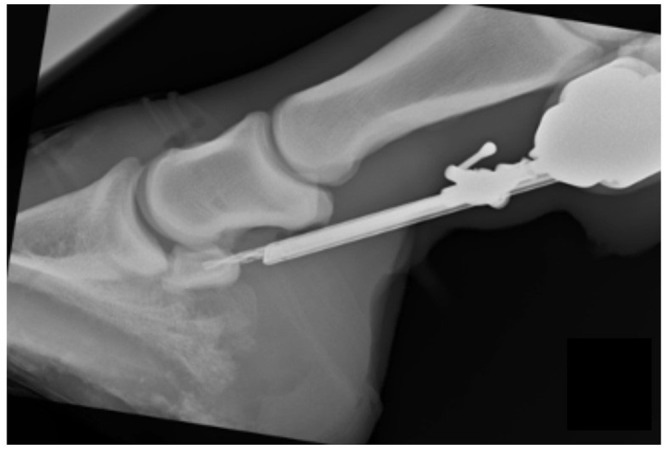
Recently, Bo Brock, DVM, DABVP (equine), from Brock Veterinary Clinic, in Lamesa, Texas, collaborated with colleagues from the College of Veterinary Medicine in Georgia, Colorado State University, and Texas A&M University to evaluate a procedure called core osteostixis. The rationale for this study was that many horses with medullary sclerosis and osseous cystlike lesions of the navicular bone fail to respond to podiatry and/or medical management.
“Osteostixis involves drilling through the cortex of the bone to the medullary cavity,” explains Brock. “It decompresses the medullary cavity. Increased pressure intramedullary may decrease blood flow through the sclerotic cortex.”
Brock et al. conducted a randomized, double-blind, self-controlled trial of seven client-owned horses diagnosed with bilateral podotrochlosis using standard and accepted techniques, including clinical exam, diagnostic analgesia, lack of response to medical management, and MRI. Navicular bursoscopy was performed bilaterally in all included horses, and core osteostixis was performed by drilling a single hole into the medullary cavity of the proximal portion of the navicular bone. If a cyst was present, the hole was drilled directly into the cyst whenever possible. If no cyst was present, the hole was drilled in the middle of the proximal cortex into the medullary cavity, to a depth of 75% the distance between the proximal and distal margins of the navicular bone, being certain not to penetrate the distal cortex.
They hypothesized that the limb with core osteostixis would have a significantly greater reduction in lameness than the contralateral limb that had bursoscopy only. In their study, Brock’s team observed significant improvement in lameness in forelimbs undergoing core osteostixis, suggesting this surgical procedure improved outcomes. Five of the seven limbs in the osteostixis group were sound at 24 weeks compared with only one of seven limbs in the bursoscopy group. On follow-up, MRI showed five of the seven horses that had cystlike lesions bilaterally prior to surgery still had cysts following therapy that remained unchanged. Further, new DDFT tears were identified in three of the seven limbs that underwent core osteostixis.
“I have been performing this surgery for 10 years now and have showed other veterinarians how to do it,” says Brock. “It is technically demanding, which may limit the number of surgeons offering this service, but there are several referral practices and veterinary schools that do.”
Concluding Remarks
Podotrochlosis negatively affects many horses’ performance and quality of life, and treatment options leave much to be desired.
“While we cannot conquer this degenerative disease, we may be able to reduce its severity somewhat,” says Fürst.
He says four specific actions might help reduce the incidence and severity of podotrochlosis:
1. Breeding healthy horses due to the presumptive genetic component.
2. Focusing on hoof care during the rearing phase.
3. Using proper hoof protection as adults.
4. Training and handling horses carefully.
Related Reading
- Horse Hoof Changes Over an 8-Week Growth Period
- Tips and Tricks to Acquiring Great Equine Foot Radiographs
- 10 Radiographic Prepurchase Abnormalities in Sport Horses
Stay in the know! Sign up for EquiManagement’s FREE weekly newsletters to get the latest equine research, disease alerts, and vet practice updates delivered straight to your inbox.
![[Aggregator] Downloaded image for imported item #19769](https://s3.amazonaws.com/wp-s3-equimanagement.com/wp-content/uploads/2026/05/31141823/EDCC-Unbranded-11-scaled-1-768x512.jpeg)

