Superficial digital flexor tendon (SDFT) injuries usually heal by fibrosis, said Roger Smith, MA, VEPMB, PHD, FHEA, DECVS, FRCVS, at the 2022 BEVA Equine Conference in Britain. Pain and lameness are a feature in some SDFT injuries, but not necessarily all. However, many individuals do experience prolonged lameness. Different stages of healing require different approaches. Smith stressed that it is necessary to fine-tune and tailor therapy rather than applying a one-size-fits-all strategy.
Mitigating Inflammation
Tendons heal in three overlapping phases: inflammation, proliferation and remodeling. The quality of healing is variable between individuals. The gold standard of care that addresses the acute phase of an SDFT injury is 80% of the battle, Smith reported. While decreasing inflammation is important, he stressed that inflammation is designed to removed damaged tissue. Therefore, mitigating inflammation is good, but you should not mitigate so much as to completely eliminate the inflammatory response. Anti-inflammatory protocols include physical therapy; cryotherapy; compressive bandaging to minimize edema; fetlock joint support and rest; reducing the progression of pathology through immobilization with casts or splints (in some cases); and pharmaceutical drugs like short-acting corticosteroids (although these could interfere with healing) or NSAIDs for analgesic effects. The objective is to decrease natural inflammation so that it doesn’t persist past the acute phase of injury.
Physical Rehabilitation
In the subacute or chronic phase of superficial digital flexor tendon injuries, the gold standard of care incorporates physical rehabilitation. This is critical to promote fibroplasia, optimize organization of the scar, promote remodeling and prevent re-injury. Walking is gradually increased with progression to trot after 4-5 months. Smith advised that a key to success relies on ultrasound monitoring every three months or after any change in exercise level. Ideally, one can visualize improved echogenicity and fiber alignment and achieve a decrease (or at least no change) in a lesion’s cross-sectional area to less than 10%. A doppler signal, which is a surrogate marker of inflammation, might increase initially and then decrease in later stages of healing.
Non-Invasive Monitoring
Other non-invasive monitoring options have demonstrated a linear relationship between force through the limb and the angle of the fetlock. Limb stiffness in the injured leg can be compared to the contralateral limb to track progress. Records of the extension of the fetlock when loaded and unloaded further help with monitoring.
Injections
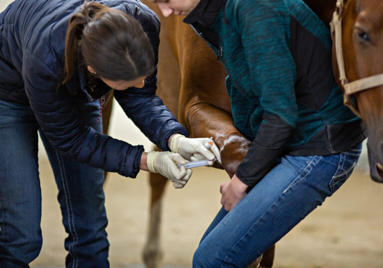
Healing improvement can be achieved with intralesional injection of PRP or MSCs, especially because an injury provides a receptacle for cells. The objective is to regenerate tissue to restore function and/or improve organization of the tendon fibers. MSCs have the potential to improve the mechanics, organization and composition of tendon fibers while also halving the risk of re-injury. At this time, PRP has very limited clinical evidence for rapid filling in of suspensory ligament lesions. IRAP is associated with significant decreases in lameness. (An alpha-2-macroglobulin applied in the acute phase shows no evidence of efficacy, Smith added.)
Smith remarked that laser treatment studies have not consistently used control groups. Therefore, while safe, there is no evidence of efficacy. A lesion might decrease in size and improve vascularity from the laser-induced heat, but it might result in a crimp pattern of the fibers.


