This article originally appeared in the Fall 2026 issue of EquiManagement. Sign up here for a FREE subscription to EquiManagement’s quarterly digital or print magazine and any special issues.
Primary back pain can stem from bone, joint, ligament, tendon, muscle, or any combination of these structures anywhere along the length of the horse’s body. As such, it’s a complicated condition to diagnose and manage. Additionally, clinical signs of back pain can be highly variable and nonspecific, including “poor performance” and “behavior problems.” Other reported clinical signs of back pain include:
- Bruxism.
- Tail swishing/altered tail carriage.
- Altered gait such as bunny hopping or lameness.
- Difficulty sliding/stopping, holding canter leads, bending.
- Missing lead changes.
- Unwilling to go forward and refusing jumps.
- Loss of topline/muscle atrophy.
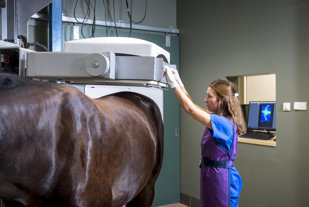
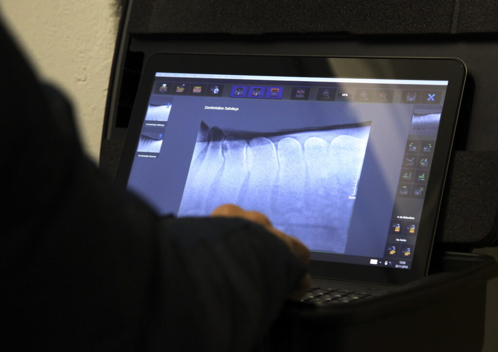
Radiology and ultrasonography are commonly the first diagnostic tools practitioners reach for upon suspicion of back pain. Additional modalities might include nuclear scintigraphy, thermography, and computed tomography (CT) based on availability, owner’s financial ability, and practitioner’s preference.
Once practitioners have those images, though, what do we do with that information? More specifically, which changes are clinically significant?
“The difficulty with image interpretation comes from the extreme variability in clinical relevance when comparing different horses that have similar findings,” said Natasha Werpy, DVM, DACVR, of Equine Diagnostic Imaging, in Archer, Florida, during a presentation at the 2024 AAEP Convention. “The correlation between imaging abnormalities and clinical examination is always crucial for determining clinical relevance. However, in the back it becomes even more critical. The clinical examination must be more heavily relied upon due to the variable clinical relevance of structural abnormalities that can be identified with back imaging.”
Henderson et al. (2024) echoed this sentiment, noting, “The past few years have seen a surge of reports on the presence of anatomical variations affecting the equine distal cervical vertebral column, providing mixed conclusions as to whether these variations are associated with clinical signs of neck dysfunction.”
“Several studies have reported a wide range of radiographic and scintigraphic changes to be present without corresponding clinical symptoms, and vice versa,” adds Lynne Harrison, MSc, PhD candidate at the University of Queensland, Australia. “Radiographic and scintigraphic findings need to be taken into context with clinical findings for accurate diagnosis and treatment planning.”
In this article, we’ll present common conditions of the equine neck and back, typical imaging findings, and whether they correlate with clinical disease.
How Common Is Back Pain in Horses?
Marshall-Gibson et al. (2023) conducted a survey of equine veterinarians about trends in diagnosing and managing back pain. They reported that back pain occurs in almost all (94%) ridden horses, due to either primary or secondary causes. Practitioners consider kissing spines (close, impinging, or overriding dorsal spinous processes) the most common primary cause of back pain. Runners-up include degenerative sacroiliac joint disease and osteoarthritis (OA) of the lumbar and thoracic articular process joints.
Additional causes might include ventral spondylosis, intervertebral disc disease, vertebral fractures, desmopathy of the supraspinous ligament or intraspinous ligament, and longissimus muscle strain.
Dorsal Spinous Process Pathology
The thoracic vertebrae can typically be radiographed with relative ease and spinous process fractures readily identified. In contrast, the clinical relevance of the radiographic findings in spinous process cases that are close, impinging, or overriding are not as easily interpreted.
“There are four grades that we use for classifying different severities of overriding dorsal spinous processes based on Dyson and Denoix (2011),” says José M. García-López, VMD, DACVS, DACVSMR, associate professor of large animal surgery at the University of Pennsylvania’s New Bolton Center. “While this grading system provides a useful framework for describing radiographic severity, radiographic grade alone does not necessarily correlate with clinical pain or poor performance. Clinical signs, physical examination findings, diagnostic analgesia, and additional imaging modalities should be considered when determining the clinical significance of overriding dorsal spinous process (ORDSP) lesions.”
Further, Werpy says the literature suggests there are “numerous radiographic abnormalities of the spinous processes of asymptomatic horses.”
García-López concurs, adding “Lysis and sclerosis may also be noted in an asymptomatic horse. For that reason, the presence of radiographic changes alone should not be used to conclude that a horse is suffering from clinical ORDSP. ”
Dorsal spinous process issues can also be noted in the lumbar region, although less frequently than the thoracic region.
“Imaging the thoracolumbar spine can be challenging due the size and anatomy of the horse, and imaging requires powerful enough equipment to penetrate the dense tissues to obtain good-quality images,” says Harrison.
She suggests using X-ray equipment with an output of 150 kV and mA of 250-500. She also recommends practitioners direct the X-ray beam perpendicular to the vertebral canal to obtain diagnostic images of the equine spine and DSPs. “Further, obtaining ventral oblique images at a 20-degree angle can be beneficial for assessing the left and right thoracic synovial intervertebral articulations.”
OA of the Articular Facets
García-López’s preference for assessing articular facet OA is CT, which allows for a three-dimensional interpretation of the process joints that cannot be completely assessed with X-rays or ultrasound.
If using X-rays to diagnose OA of the facets, García-López suggests a minimum of four to five views, including lateral, right 45-degree proximal to left distal, and left 45-degree proximal to right distal views to visualize the facet joint.
“If a practitioner can get a good projection with enough definition, you can see bony remodeling shown as periarticular osteophyte (bone spur) formation,” says García-López.
Harrison adds that ultrasonography is an excellent tool for diagnosing OA of the facet joints. She suggests clipping the site of interest and using a microconvex or linear probe to assess the facet joints.
As noted by Henderson et al., “Although many horses with neck stiffness or pain have osteoarthropathy of one or more cervical articular process joints and treatment of affected joints leads to improvement in clinical signs, studies have now shown that the presence of articular process joint osteoarthropathy is common and often not associated with clinical signs.”
Further, García-López says normal variations in the anatomy can occur, potentially complicating the clinical picture.
“This is one of the challenges we have, as we not only have normal variations but also variations that are truly abnormal but not clinically relevant,” he adds.
Ventral Spondylosis
Veterinarians frequently observe osseous proliferation of the ventral vertebral margins, considering it an incidental finding. However, if an equine athlete has persistent performance limitations after ruling out all other causes of back pain, then it’s worth examining the impact of spondylosis further.
“In some cases, clinical signs may be more serious when the osteophytes compress nerve roots at the intervertebral foramen or grow into the vertebral canal and compress the spinal cord,” Domańska-Kruppa et al. stated in their 2024 review article on advances in clinical diagnosis of back pain.
Intervertebral disc spaces can be assessed for either narrowing or collapse, notes Werpy, and CT and scintigraphy might be useful tools for this purpose.
Transposition of Ventral Lamina of C6 onto C7 (TC67)
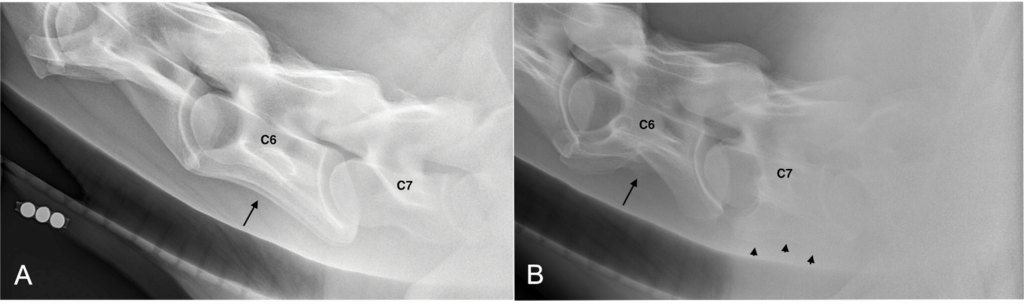
An increasingly hot topic in equine practice is the clinical relevance of TC67. With this condition, the ventral lamina of the transverse process of the sixth cervical vertebra is transposed to the ventral aspect of the transverse process of the seventh cervical vertebra. It can occur uni- or bilaterally.
Henderson et al. retrospectively reviewed medical records and radiographs of horses that had X-rays of the cervical necks performed between August 2020 and August 2023. In total, 135 horses were imaged and 27 (20%) were diagnosed with TC67, making this a relatively common diagnosis.
In terms of clinical relevance, however, signs of neurologic disease occurred equally in horses with and without TC67.
Close to twice as many horses (19%) were ultimately diagnosed with neck pain when compared to the group without TC67 (10%); however, this did not reach statistical significance (P = .68). When only looking at the group of horses diagnosed with neck pain, 5 (31%) were found to have TC67 and 11 (69%) did not.
“Although the number of horses is small in these groups, this finding was significantly different from the horses for which neck pain was not reported, in which TC67 occurred in 22 (18%) and did not occur in 97 (82%; P < .001),” wrote the researchers.
In conclusion, they wrote, “Our data suggested that TC67 is more common in horses with neck pain; however, these results should be interpreted with caution. Our findings added to the literature on clinical signs of neck dysfunction and did not show an association between TC67 and neurologic disease but did highlight the need for further study of horses with signs of neck pain.”
Sacroiliac (SI) Pain
According to the 2023 survey conducted by Marshall-Gibson et al., degeneration of the sacroiliac joint occurs commonly. García-López (2018) found that of 39 lameness cases, 20 horses had sacroiliac pain and/or region abnormalities and 100% of racehorses had degeneration of the SI joint observed at the time of euthanasia. Two common causes of SI pain include OA of the SI joint and pathology of the dorsal sacroiliac ligament, which is a bilateral structure. Ultrasonography is the diagnostic tool of choice, typically rectally. Clinically relevant findings include enlarged hypoechoic dorsal sacroiliac ligaments with fiber disruption and unilateral decrease in area in cases of chronic desmitis.
“The primary change that worries me is significant osteophyte formation on the ventral aspect of the SI joints as seen on ultrasound per rectum,” says García-López. “Use caution not to overinterpret bilaterally symmetric hypoechoic lesions of the dorsal spinous ligament.”
Integrating Neck and Back Imaging Into the Prepurchase Examination
Given that some imaging findings don’t necessarily have clinical relevance, should images of the neck or back be included in prepurchase exams?
“This is always a tough question to answer based on the incorrect perceptions of people when they see an ‘ugly’ back,” García-López says. “I discuss with potential buyers what I am seeing on radiographs in combination with my evaluation, level of fitness/muscling of the horse in the epaxial (back) region, purpose, and what has been done in the past in order to try to give them the best possible guidance. I explain that X-rays alone are not worthless, but we cannot make conclusions based on X-rays alone due to the high number of normal horses with abnormal radiographs of this region.”
He adds that if the buyer plans to sell the horse in the future, they might have issues with other veterinarians or potential buyers not considering or “flunking” the horse based on those films, even if the area has not been a problem.
Harrison adds that many purchasers choose not to take radiographs if the horse displays no pain or dysfunction on assessment.
“For owners that do undertake imaging, if there is evidence of overriding dorsal spinous processes, for example, and the horse is asymptomatic at the time of exam, it is impossible to say that the horse will develop pain going forward,” she says. “However, the purchaser will be informed and can decide based on the findings and recommendations of the veterinarian. If the horse moves well and shows no signs of pain, the findings may just be incidental. It is not black and white unfortunately.”
Increasing Use of CT for the Caudal Spine
Nadine Ogden, BVSc, CertAVP(EL), MScVPS, FHEA, DACVSMR, MRCVS, American & RCVS Specialist in Equine Sports Medicine and Rehabilitation from B&W Equine Hospital in Berkeley, U.K., recently reported that large-bore CT units could reliably produce diagnostic images of the caudal spine and pelvis (Ogden et al. 2024).
“Anatomic variations were common, such as the location of the widest interspinous space, shape of the last lumbar vertebra, and degree of fusion/modeling,” she says.
In a follow-up study also published in 2024, Ogden et al. reported on pathologic lesions identified in 56 horses undergoing CT of the caudal spine and pelvis while anesthetized. These horses presented primarily for lameness (n=30) and poor performance (n=18).
Osteoarthritis of the SI joint was diagnosed in 28 horses, pathology of the intervertebral disc joint in 12 horses, pelvic fracture in nine horses, and ventral spondylosis in six.
“This is a retrospective case series, so in many cases a definitive causal relationship between CT findings and clinical signs cannot be stated with certainty,” Ogden explains. “What CT gave us was diagnostic clarity we hadn’t previously had: We could see lesions that would have been missed or undercharacterized on scintigraphy, radiography, or ultrasound. Whether a lesion is the primary driver of clinical signs requires integration of the full clinical picture, such as response to local anesthesia or treatment, the horse’s history, gait analysis, and clinician judgment.”
Concluding Remarks
Widespread availability and use of imaging modalities allows practitioners to image essentially all regions of the equine spinal column. However, not all imaging “abnormalities” have clinical relevance. Sifting through the array of incidental findings in the face of a horse with back pain proves clinically challenging.
“Radiographs alone should not decide whether a horse has clinical ORDSP,” says García-López. “We always need to put it into context including athletic discipline, the physical examination and clinical evaluation, past history of the horse, and ability of the rider to have the best possible picture.”
Harrison adds, “When imaging is used in isolation, a full picture of the problem may be difficult to establish, leading to inappropriate treatment and unsatisfactory outcomes.”
Related Reading
- Coordinating Care for the Modern Horse
- Laser Acupuncture vs. Electroacupuncture for Equine Back Pain
- Success Rates of Different Kissing Spines Management Options
Stay in the know! Sign up for EquiManagement’s FREE weekly newsletters to get the latest equine research, disease alerts, and vet practice updates delivered straight to your inbox.